Observational Study
. 2022 Jun 23;12(1):10714.
doi: 10.1038/s41598-022-14663-3.

Canine root/cortical bone relation (CRCR) and the orthodontic tooth movement

Affiliations
Observational Study

Canine root/cortical bone relation (CRCR) and the orthodontic tooth movement

Amr R El-Beialy et al. Sci Rep. .

Abstract

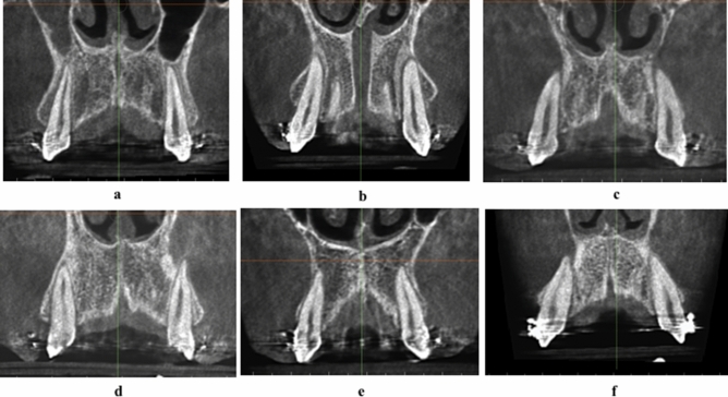
This observational study evaluates three-dimensionally the relation between the root of maxillary canine and overlying labial cortical plate of bone during orthodontic canine retraction. Eighty-four bilateral maxillary canines in 42 patients were retracted in the extraction space of first premolars, using conventional orthodontic NiTi retraction spring delivering 150gm. Three-dimensional evaluation at the cusp tip, root apex, and the overlying cortical bone was done based on Classification of Root/Cortical bone relation (CRCR) before and after canine retraction. 168 observations of the canines pre- and post-retraction showed a mean distal movement of the canine cusp tip of 3.78(± 2.05) mm, while the canine root apex was almost stationary. Scarcely, 5.4% of the canine roots and root apices persisted in the medullary bone during retraction, while 16.1% contacted the overlying cortical bone. Fenestration of the overlying cortical bone by the canine roots or root apices occurred in 78.6% of the sample. The unembellished intimacy between the canine root and apex to the overlying thick dense cortical bone might have the decelerating effect on the maxillary canine retraction. The natural bone plate labial to the maxillary canine root did not yield infront nor enlarge due to canine retraction, but else defeated the current orthodontic biomechanical implementation.


Conflict of interest statement

The authors declare no competing interests.

Figures

Figure 1
Figure 1
(a) Maxillary canine retraction using conventional mechanics, (b) maxillary arch CBCT before canine retraction, (c) maxillary arch CBCT after canine retraction, (d) distances traveled by canine cusp tip and root apex on CBCT, (e) superimposed CBCTs before, and after canine retraction showing the amount of canine tipping.
Figure 2
Figure 2
(a) CRCR Grade A: the canine root and root apex totally present inside the medullary bone leaving a continued thickness of the overlying cortical bone. (b) CRCR Grade B: the canine roots or root apices in contact with the overlying cortical bone, with thinning of the overlying cortical bone. (c) CRCR Grade C: penetration of the canine roots of the overlying cortical bone, and loss of the continuity of the cortical bone (fenestration or deep dehiscence). (d) Proportional percentage of each of CRCR grades.
Figure 3
Figure 3
Examples of CRCR Grade A.
Figure 4
Figure 4
Examples of CRCR Grade B.
Figure 5
Figure 5
Examples of CRCR Grade C.
Figure 6
Figure 6
(a,b) Canines of the same patients in Grade A in the pre-retraction position (a) changed to grade C in the post-retraction position (b), (c–f) canines of the same patients in Grade B in the pre-retraction position (c,e) changed to grade C in the post-retraction position (d,f), (g–j) canines of the same patients in Grade C in the pre-retraction position(g,i) remained as grade C in the post-retraction position (h,j).
Figure 7
Figure 7
(a) Identical grade A of CRCR bilaterally. (b) Identical grade B of CRCR bilaterally. (c) Identical grade C of CRCR bilaterally. (d) Different grades of CRCR bilaterally (Right grade A/Left grade C). (e) Different grades of CRCR bilaterally (Right grade B/Left grade A). (f) Different grades of CRCR bilaterally (Right grade C/Left grade B).
Figure 8
Figure 8
CBCT volumes examined in the 3D solid bone view.

References

    1. Masella RS, Meister M. Current concepts in the biology of orthodontic tooth movement. Am. J. Orthod. Dentofac. Orthop. 2006;129:458–468. doi: 10.1016/j.ajodo.2005.12.013. - DOI - PubMed
    1. Krishnan V, Davidovitch Z. Cellular, molecular, and tissue-level reactions to orthodontic force. Am. J. Orthod. Dentofacial Orthop. 2006;129(469):e1–32. - PubMed
    1. Proffit, W. R. & Fields, H. W. Contemporary Orthodontics. (2000).
    1. Jäger F, Mah JK, Bumann A. Peridental bone changes after orthodontic tooth movement with fixed appliances: A cone-beam computed tomographic study. Angle Orthod. 2017;87:672–680. doi: 10.2319/102716-774.1. - DOI - PMC - PubMed
    1. Tweed CH. A philosophy of orthodontic treatment. Am. J. Orthod. Oral Surg. 1945;31:74–103. doi: 10.1016/S0096-6347(45)90163-3. - DOI

Publication types