Review
. 2022 Aug;29(8):1433-1449.
doi: 10.1038/s41418-022-01028-6. Epub 2022 Jun 23.

Past, present, and future perspectives of transcription factor EB (TFEB): mechanisms of regulation and association with disease

Affiliations
Review

Past, present, and future perspectives of transcription factor EB (TFEB): mechanisms of regulation and association with disease

Anderson Tan et al. Cell Death Differ. 2022 Aug.

Abstract

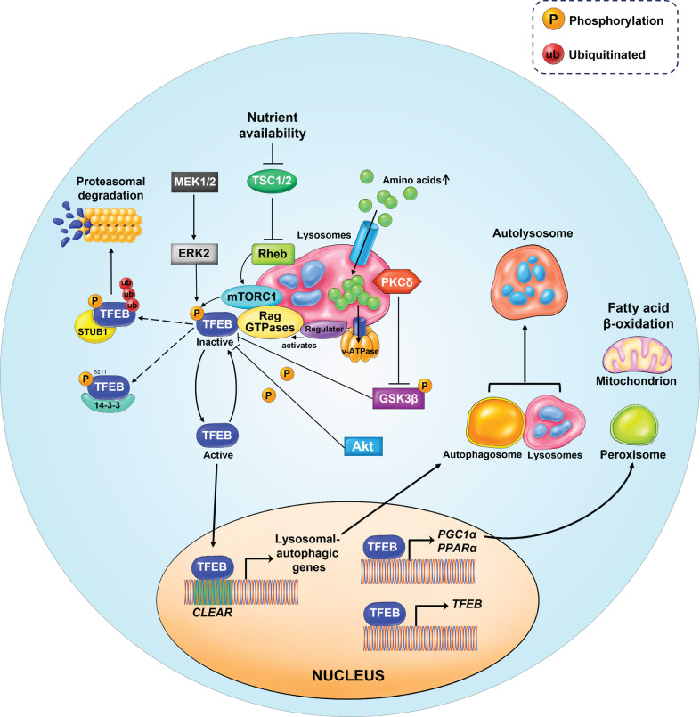
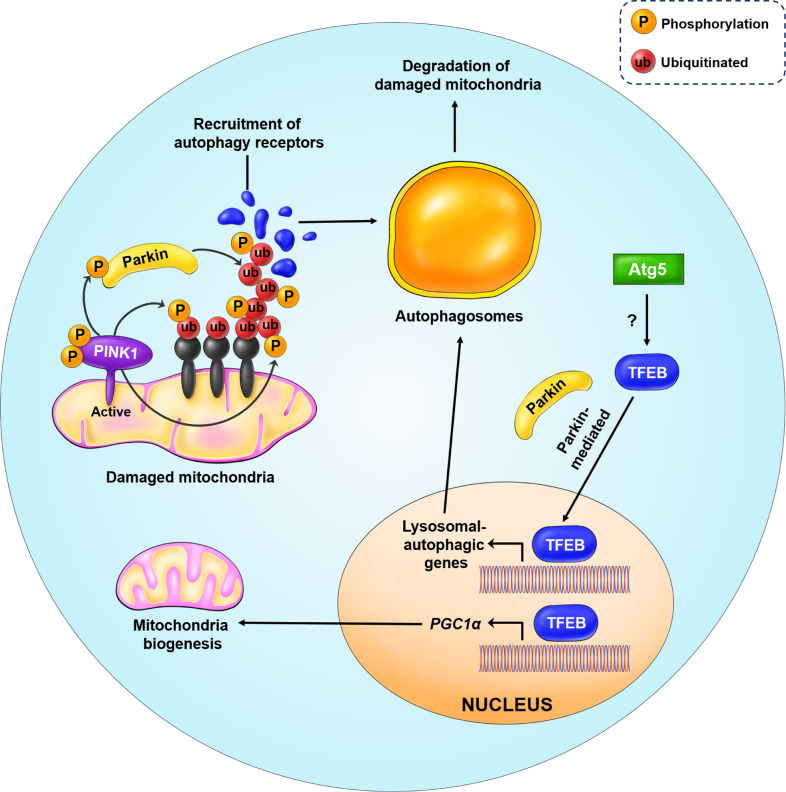
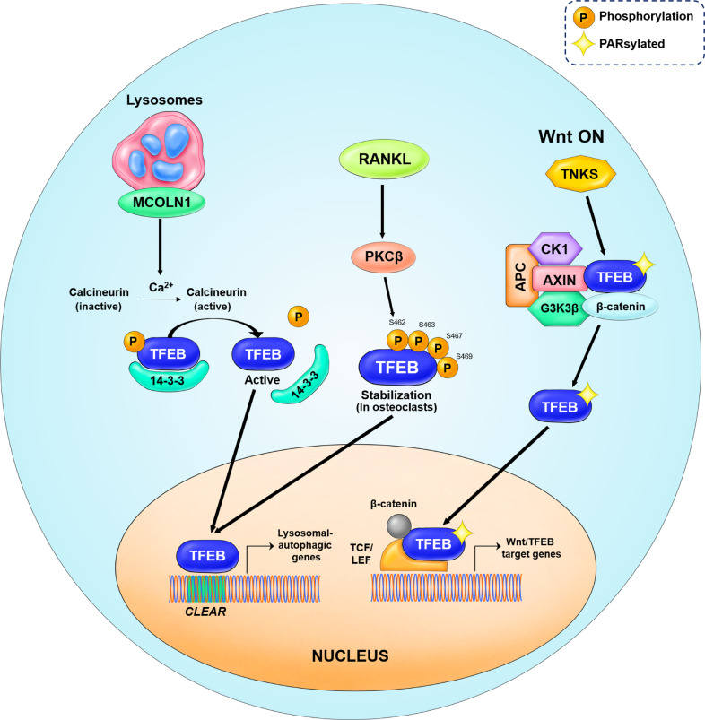
Transcription factor EB (TFEB), a member of the MiT/TFE family of basic helix-loop-helix leucine zipper transcription factors, is an established central regulator of the autophagy/lysosomal-to-nucleus signaling pathway. Originally described as an oncogene, TFEB is now widely known as a regulator of various processes, such as energy homeostasis, stress response, metabolism, and autophagy-lysosomal biogenesis because of its extensive involvement in various signaling pathways, such as mTORC1, Wnt, calcium, and AKT signaling pathways. TFEB is also implicated in various human diseases, such as lysosomal storage disorders, neurodegenerative diseases, cancers, and metabolic disorders. In this review, we present an overview of the major advances in TFEB research over the past 30 years, since its description in 1990. This review also discusses the recently discovered regulatory mechanisms of TFEB and their implications for human diseases. We also summarize the moonlighting functions of TFEB and discuss future research directions and unanswered questions in the field. Overall, this review provides insight into our understanding of TFEB as a major molecular player in human health, which will take us one step closer to promoting TFEB from basic research into clinical and regenerative applications.


Conflict of interest statement

The authors declare no competing interests.

Figures

Fig. 1
Fig. 1. Domain structure of MiT/TFE proteins and their homologs in primitive metazoans.
A The MiT/TFE family comprises of four members: TFEB, MITF, TFE3, and TFEC, all of which share conserved basic helix-loop-helix (bHLH) and leucine zipper (Zip) domains. B Different species harbor different MiT/TFE members owing to variation in whole-genome duplication. C The CLEAR motif, which can be found in the majority of autophagy-lysosomal promoters, is recognized by MiT/TFE proteins. Abbreviations for each domain are as follows: activation domain (AD), glutamine-rich (Gln rich), proline-rich (Pro rich), serine-rich (Ser rich), and proline + arginine (Pro + Arg) domain.
Fig. 2
Fig. 2. Pathophysiological functions and importance of the TFEB protein.
TFEB regulates the lysosomal biogenesis & autophagy pathway, which is crucial for proper tissue-specific regulation and TFEB-mediated intracellular clearance of toxic protein aggregates (upper panel). The number of research articles has increased along with the interest in and perceived importance of the TFEB protein (bottom panel).
Fig. 3
Fig. 3. Timeline of the major advances in TFEB research, in the 30 years since discovery.
The timeline summarizes 30 years of major advances and key findings in TFEB research, from 1990 through to 2021.
Fig. 4
Fig. 4. Phosphorylation sites in the TFEB domain structure and their regulatory role.
TFEB can be phosphorylated at multiple serine residues by upstream kinases, such as mTOR, GSK3β, ERK2, AKT, and PKCβ. Phosphorylation of TFEB by PKCβ promotes TFEB protein stabilization. Moreover, functional studies show that TFEB localization to the lysosomal membrane is important for its inhibition by mTORC1 complex-mediated phosphorylation.
Fig. 5
Fig. 5. Schematic model of TFEB upstream regulation by mTORC1.
In nutrient-rich conditions, for instance, amino stimulation, mTORC1 is recruited to the lysosomal membrane. Amino acids can freely cross the lysosomal membrane and accumulate inside the lysosomes. These amino acids are ‘sensed’ by the lysosomal lumen and signal to the Rag GTPases through the v-ATPase-Ragulator complex. Activated Rag GTPases recruit both TFEB and mTORC1 to the lysosomal membrane. Rheb promotes the activation of membrane-localized mTORC1. Active mTORC1 phosphorylates TFEB, thus causing its inactivation and inhibiting its nuclear translocation. In addition, TFEB can also be phosphorylated by ERK2, AKT, and GSK3β. Phosphorylated TFEB is recognized by STUB1, an E3-ligase, which then targets TFEB for proteasomal degradation. However, in nutrient- deprived conditions, TFEB becomes active and free from phosphorylation. Active TFEB then translocates to the nucleus, binds to the CLEAR motif, and increases the expression of genes involved in autophagy/lysosomal biogenesis. TFEB also binds to its own promoters and upregulates its expression through a self-regulatory feedback loop. TFEB is responsible for the transcriptional activation of PGC1α, the co-activator of the nuclear receptor peroxisome proliferator-activated receptor α (PPARα) and a key regulator of lipid metabolism and mitochondrial biogenesis.
Fig. 6
Fig. 6. Regulation of PINK1/Parkin-mediated mitophagy by TFEB.
In the case of mitochondria damage and loss of membrane potential, PINK1 is recruited to the OMM and activated through auto-phosphorylation. PINK1 triggers mitophagy initiation by phosphorylating the ubiquitin attached to the damaged and misfolded OMM proteins. Simultaneously, PINK1 also phosphorylates Parkin, an E3 ligase, causing its activation. Activated Parkin further amplifies the signal by further ubiquitinating misfolded OMM proteins. In addition, phosphorylated ubiquitin also recruits autophagy receptors (blue) to induce autophagosome formation, for degradation of damaged mitochondria. This process requires the transcriptional activity of nuclear TFEB to induce the expression of genes involved in autophagy biogenesis. Additionally, ATG5 activates TFEB via an unknown mechanism, while TFEB nuclear localization is Parkin-dependent. TFEB also induces the expression of PGC1α, a master regulator of mitochondrial biogenesis.
Fig. 7
Fig. 7. Schematic model of the upstream regulators of TFEB.
In starvation conditions, calcium is released from lysosomes through the mucolipin 1 (MCOLN1) channel protein. Calcium ions induce the activation of calcineurin, a phosphatase, resulting in TFEB dephosphorylation. Dephosphorylated TFEB detaches from the 14-3-3 chaperone proteins and enters the nucleus to induce autophagy-lysosomal biogenesis. PKCβ activation is mediated by RANKL. The active PKCβ phosphorylates multiple serine residues in the C-terminal region of the TFEB protein. Such phosphorylation leads to TFEB nuclear translocation, inducing lysosomal biogenesis and secretion of lysosomal hydrolases in osteoclasts. In the Wnt ON condition, TFEB undergoes the PARsylation post-translational modification. The PARsylated TFEB translocates to the nucleus to form a transcription factor complex with β-catenin-TCF/LEF1. This complex regulates a subset of genes that are distinct from the canonical β-catenin target genes, known as Wnt/TFEB target genes.

References

    1. Steingrímsson E, Copeland NG, Jenkins NA. Melanocytes and the microphthalmia transcription factor network. Annu Rev Genet. 2004;38:365–411. - PubMed
    1. La Spina M, Contreras PS, Rissone A, Meena NK, Jeong E, Martina JA. MiT/TFE family of transcription factors: an evolutionary perspective. Front Cell Dev Biol. 2020;8:609683. - PMC - PubMed
    1. Lapierre LR, De Magalhaes Filho CD, McQuary PR, Chu C-C, Visvikis O, Chang JT, et al. The TFEB orthologue HLH-30 regulates autophagy and modulates longevity in Caenorhabditis elegans. Nat Commun. 2013;4:1–8. - PMC - PubMed
    1. O’Rourke EJ, Ruvkun G. MXL-3 and HLH-30 transcriptionally link lipolysis and autophagy to nutrient availability. Nat Cell Biol. 2013;15:668–76. - PMC - PubMed
    1. Settembre C, De Cegli R, Mansueto G, Saha PK, Vetrini F, Visvikis O, et al. TFEB controls cellular lipid metabolism through a starvation-induced autoregulatory loop. Nat Cell Biol. 2013;15:647–58. - PMC - PubMed

Publication types

MeSH terms

Substances