. 2022 Jun 23;10(1):92.
doi: 10.1186/s40478-022-01393-w.

Pathological and neurophysiological outcomes of seeding human-derived tau pathology in the APP-KI NL-G-F and NL-NL mouse models of Alzheimer's Disease

Affiliations

Pathological and neurophysiological outcomes of seeding human-derived tau pathology in the APP-KI NL-G-F and NL-NL mouse models of Alzheimer's Disease

S Tok et al. Acta Neuropathol Commun. .

Abstract

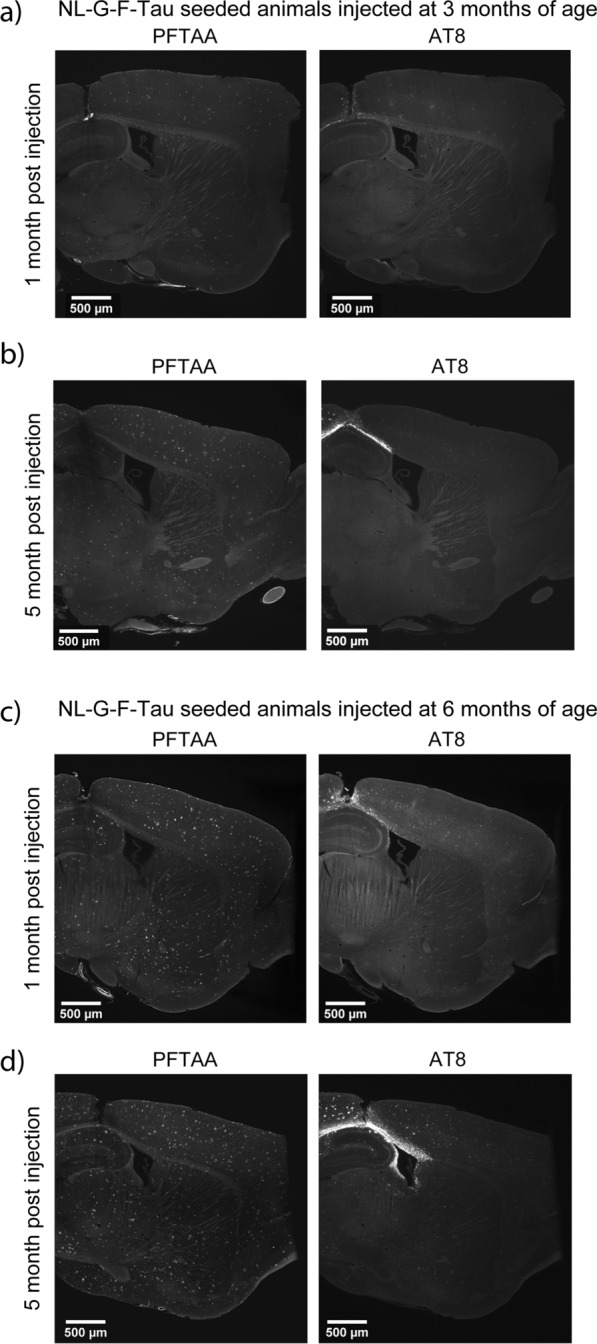
The two main histopathological hallmarks that characterize Alzheimer's Disease are the presence of amyloid plaques and neurofibrillary tangles. One of the current approaches to studying the consequences of amyloid pathology relies on the usage of transgenic animal models that incorporate the mutant humanized form of the amyloid precursor protein (hAPP), with animal models progressively developing amyloid pathology as they age. However, these mice models generally overexpress the hAPP protein to facilitate the development of amyloid pathology, which has been suggested to elicit pathological and neuropathological changes unrelated to amyloid pathology. In this current study, we characterized APP knock-in (APP-KI) animals, that do not overexpress hAPP but still develop amyloid pathology to understand the influence of protein overexpression. We also induced tau pathology via human-derived tau seeding material to understand the neurophysiological effects of amyloid and tau pathology. We report that tau-seeded APP-KI animals progressively develop tau pathology, exacerbated by the presence of amyloid pathology. Interestingly, older amyloid-bearing, tau-seeded animals exhibited more amyloid pathology in the entorhinal area, isocortex and hippocampus, but not thalamus, which appeared to correlate with impairments in gamma oscillations before seeding. Tau-seeded animals also featured immediate deficits in power spectra values and phase-amplitude indices in the hippocampus after seeding, with gamma power spectra deficits persisting in younger animals. Both deficits in hippocampal phase-amplitude coupling and gamma power differentiate tau-seeded, amyloid-positive animals from buffer controls. Based on our results, impairments in gamma oscillations appear to be strongly associated with the presence and development of amyloid and tau pathology, and may also be an indicator of neuropathology, network dysfunction, and even potential disposition to the future development of amyloid pathology.


Conflict of interest statement

The authors declare that they are working for Janssen Pharmaceutica NV. The authors declare no other commercial or financial relationships that could be construed as a potential conflict of interest.

Figures

Fig. 1
Fig. 1
Illustrated overview of the experimental study, containing the workflow for histological and electrophysiological assessment. a Timeline for both histological and electrophysiological cohorts indicating sample sizes, recording, euthanasia and injection timepoints for two age groups. B+n refers to n days after birth. D+n refers to n days after injection. b Histological workflow illustrating surgical procedure, whole-brain immunostaining, light-sheet imaging and quantification. M.p.i refers to months-post injection. c Workflow for electrophysiological assessment showing EEG recording, activity detection, and vigilance-controlled analysis readouts
Fig. 2
Fig. 2
Representative histological images from NL-G-F-Tau animals injected at 3 and 6 months of age. Representative light sheet images of amyloid (PFTAA) and AT8-positive tau pathology in 3-month-old NL-G-F-tau animals at a 1 month-post-injection and b 5 months-post-injection. Representative light sheet images of amyloid and AT8-positive tau pathology in 6-month-old NL-G-F-tau animals at c 1 month-post-injection and d 5 months-post-injection. PFTAA refers to Pentameric formyl thiophene acetic acid, AT8 refers to AT8-positive tau pathology
Fig. 3
Fig. 3
Quantification and comparisons of AT8 and amyloid pathology across genotype, and time-post-injection from animals injected at 3 or 6 months of age. a Quantification and pairwise comparisons of AT8-positive tau pathology in animals injected at 3 months of age, at 1 and 5 m.p.i. b Quantification and pairwise comparisons of AT8-positive tau pathology in in animals injected at 6 months of age, at 1 and 5 m.p.i. c Quantification and pairwise comparisons of amyloid pathology in animals injected at 3 months of age, at 1 and 5 m.p.i. d Quantification and pairwise comparisons of amyloid pathology in animals injected at 6 months of age, at 1 and 5 m.p.i. Error bars are standard error of mean. Asterisks indicate significant comparisons (p < 0.05). n.s. refers to non-significant comparisons. M.p.i refers to months-post injection
Fig. 4
Fig. 4
Power spectra quantification and comparisons between NL-G-F and NL-NL animals at baseline. Log power graphs and power spectra comparisons of NL-G-F and NL-NL animals from 1 to 50 Hz injected at a 3 months of age and b 6 months of age from the medial entorhinal cortex and retrosplenial cortex before injection. Bar plot values correspond to quantified average of blue highlighted regions. c Baseline low gamma power spectra values before tau seeding injection in 6-month-old APP-KI animals, across multiple brain regions. Error bars are standard error of mean. Asterisks indicate significant comparisons (p < 0.05)
Fig. 5
Fig. 5
Bar plots of power spectra changes and phase-amplitude coupling and in 3- and 6-month-old APP-KI animals before and 1 day after injection. Bar plots of hippocampal power values across different frequency bands in animals injected at a 3- and b 6-months of age before and 1 day after injection. c) Phase-amplitude coupling deficits immediately following injection in tau-seeded APP-KI animals at either 3 or 6 months of age. Error bars are standard error of mean. Asterisks indicate significant comparisons (p < 0.05). Pre refers to pre-injection. Post refers to 1 day after injection
Fig. 6
Fig. 6
Bar plots of longitudinal changes in power spectra and phase-amplitude coupling at 1 and 5 months-post injection. a Bar plots of hippocampal low and high gamma band power of animals seeded at 3 months of age at 1 and 5 months-post-injection. b Bar plots of hippocampal low and high gamma band power of animals seeded at 6 months of age at 1 and 5 months-post-injection. Theta1-low gamma and Theta-2 low gamma phase-amplitude coupling comparisons in the hippocampus c of animals seeded at 3 months of age and d of animals seeded at 6 months of age at 5 m.p.i. e Comparison of low gamma power in the retrosplenial cortex between of animals seeded at 3 or 6 months of age at 1 and 5 months-post injection. Error bars are standard error of mean. Asterisks indicate significant comparisons (p < 0.05). M.p.i refers to months-post injection

References

    1. Thal DR, Rüb U, Orantes M, Braak H. Phases of A beta-deposition in the human brain and its relevance for the development of AD. Neurology. 2002;58:1791–1800. - PubMed
    1. Braak H, Alafuzoff I, Arzberger T, Kretzschmar H, Del Tredici K. Staging of Alzheimer disease-associated neurofibrillary pathology using paraffin sections and immunocytochemistry. Acta Neuropathol. 2006;112:389–404. - PMC - PubMed
    1. Hall AM, Roberson ED. Mouse models of Alzheimer’s disease. Brain Res Bull. 2012;88:3–12. - PMC - PubMed
    1. Radde R, et al. Abeta42-driven cerebral amyloidosis in transgenic mice reveals early and robust pathology. EMBO Rep. 2006;7:940–946. - PMC - PubMed
    1. Yoshiyama Y, et al. Synapse loss and microglial activation precede tangles in a P301S tauopathy mouse model. Neuron. 2007;53:337–351. - PubMed

Publication types