. 2022 May 27;11(6):1070.
doi: 10.3390/antiox11061070.

Alanyl-Glutamine Protects against Lipopolysaccharide-Induced Liver Injury in Mice via Alleviating Oxidative Stress, Inhibiting Inflammation, and Regulating Autophagy

Affiliations

Alanyl-Glutamine Protects against Lipopolysaccharide-Induced Liver Injury in Mice via Alleviating Oxidative Stress, Inhibiting Inflammation, and Regulating Autophagy

Jiaji Hu et al. Antioxidants (Basel). .

Abstract

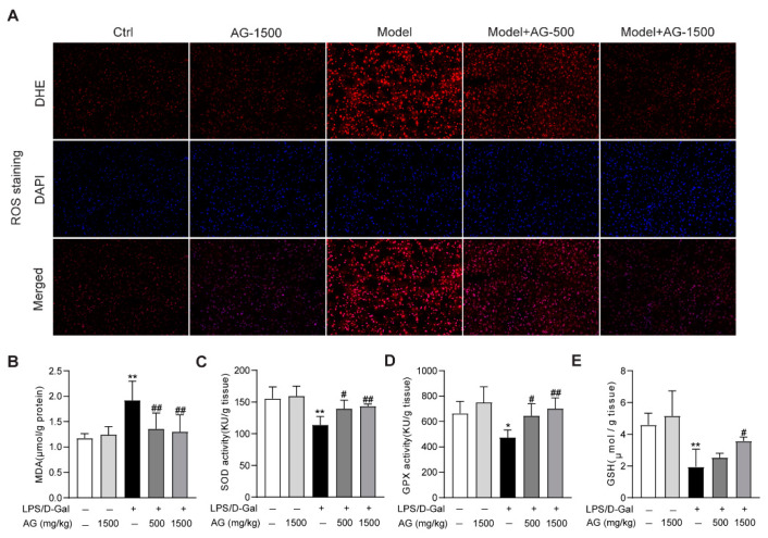
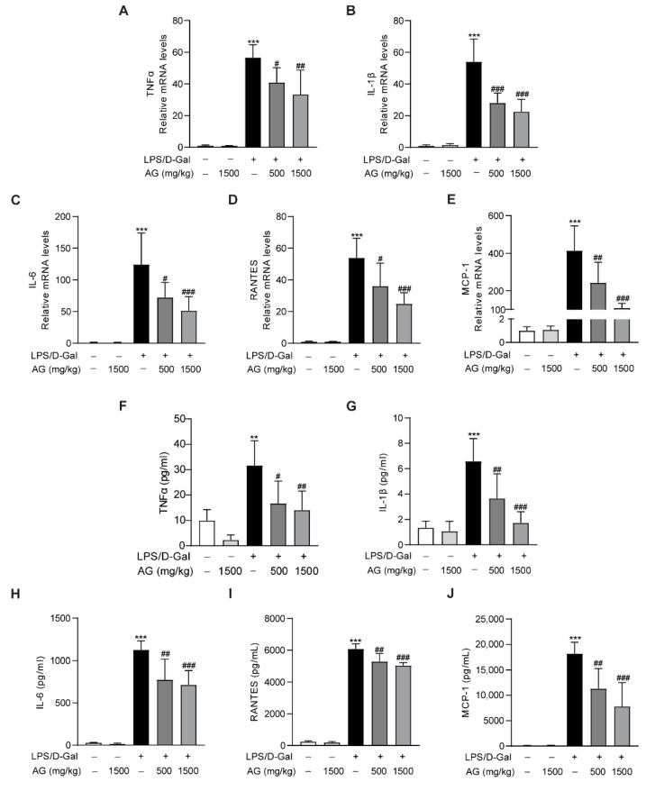
Acute liver injury is a worldwide problem with a high rate of morbidity and mortality, and effective pharmacological therapies are still urgently needed. Alanyl-glutamine (Ala-Gln), a dipeptide formed from L-alanine and L-glutamine, is known as a protective compound that is involved in various tissue injuries, but there are limited reports regarding the effects of Ala-Gln in acute liver injury. This present study aimed to investigate the protective effects of Ala-Gln in lipopolysaccharide (LPS)-induced acute liver injury in mice, with a focus on inflammatory responses and oxidative stress. The acute liver injury induced using LPS (50 μg/kg) and D-galactosamine (D-Gal) (400 mg/kg) stimulation in mice was significantly attenuated after Ala-Gln treatment (500 and 1500 mg/kg), as evidenced by reduced plasma alanine transaminase (ALT) (p < 0.01, p < 0.001), aspartate transaminase (AST) (p < 0.05, p < 0.001), and lactate dehydrogenase (LDH) (p < 0.01, p < 0.001) levels, and accompanied by improved histopathological changes. In addition, LPS/D-Gal-induced hepatic apoptosis was also alleviated by Ala-Gln administration, as shown by a greatly decreased ratio of TUNEL-positive hepatocytes, from approximately 10% to 2%, and markedly reduced protein levels of cleaved caspase-3 (p < 0.05, p < 0.001) in liver. Moreover, we found that LPS/D-Gal-triggered oxidative stress was suppressed after Ala-Gln treatment, the effect of which might be dependent on the elevation of SOD and GPX activities, and on GSH levels in liver. Interestingly, we observed that Ala-Gln clearly inhibited LPS/D-Gal exposure-induced macrophage accumulation and the production of proinflammatory factors in the liver. Furthermore, Ala-Gln greatly regulated autophagy in the liver in LPS/D-Gal-treated mice. Using RAW264.7 cells, we confirmed the anti-inflammatory role of Ala-Gln-targeting macrophages.

Keywords: acute liver injury; alanyl-glutamine; apoptosis; inflammation; oxidative stress.


Conflict of interest statement

The authors declare no conflict of interest.

Figures

Figure 1
Figure 1
Effects of Ala-Gln on LPS/D-Gal-induced hepatic pathological and biochemical parameters in mice. (A) Liver tissues were stained with HE for histopathological analysis (original magnifications, ×400), and the degree of liver injury was scored. (BD) plasma ALT, AST, and LDH levels. Data A is presented as medians with interquartile range. Data B–D are expressed as mean ± SD; n = 6–8 in each group. *** p < 0.001 vs. vehicle-treated control group, ## p < 0.01, ### p < 0.001 vs. LPS/D-Gal-treated model group.
Figure 2
Figure 2
Ala-Gln suppressed apoptosis in LPS/D-Gal-treated mice. (A) TUNEL staining (original magnification, ×400) of liver tissues. (B) Quantitation of TUNEL-positive cells in liver. (C,D) Protein expression levels of cleaved-caspase-3 in liver. Data are expressed as mean ± SD, n = 6–8 in each group. *** p < 0.001 vs. vehicle-treated control group, # p < 0.05, ### p < 0.001 vs. LPS/D-Gal-treated model group.
Figure 3
Figure 3
Antioxidant effects of Ala-Gln in liver tissues from mice that received LPS/D-Gal injection. (A) ROS detection via DHE staining (original magnification, ×200) in liver tissues. (B) Hepatic MDA levels. (C) Hepatic SOD activities. (D) Hepatic GPX activities. (E) Hepatic GSH levels. Data are expressed as mean ± SD, n = 6–8 in each group. * p < 0.05, ** p < 0.01 vs. vehicle-treated control group, # p < 0.05, ## p < 0.01 vs. LPS/D-Gal-treated model group.
Figure 4
Figure 4
IHC analysis results showing the levels of F4/80 and CD68 in liver tissues in mice. (A) IHC staining of F4/80 of liver tissues (original magnifications, ×400), and quantitation of F4/80 positive area. (B) IHC staining of CD68 of liver tissues (original magnifications, ×400), and quantitation of CD68 positive area. Data are expressed as mean ± SD, n = 6–8 in each group. *** p < 0.001 vs. vehicle-treated control group, # p < 0.05, ## p < 0.01, ### p < 0.001 vs. LPS/D-Gal-treated model group.
Figure 5
Figure 5
Impact of Ala-Gln on LPS/D-Gal-induced inflammatory responses in liver and plasma. (AE) mRNA expression levels of TNF-α, IL-1β, IL-6, RANTES, and MCP-1 in liver tissues. (FJ) Protein levels of TNF-α, IL-1β, IL-6, RANTES, and MCP-1 in plasma. Data are expressed as mean ± SD, n = 6–8 in each group. ** p < 0.01, *** p < 0.001 vs. vehicle-treated control group, # p < 0.05, ## p < 0.01, ### p < 0.001 vs. LPS/D-Gal-treated model group.
Figure 6
Figure 6
Effect of Ala-Gln on autophagy in liver in LPS/D-Gal-treated mice. (A) Protein expression levels of p62, Beclin-1, ATG-7, and LC3B in liver were assessed using Western blot. (BE) Quantitation of p62, Beclin-1, ATG-7, and LC3B-II/I expression. Data are expressed as mean ± SD, n = 6–8 in each group. ** p < 0.01, *** p < 0.001 vs. vehicle-treated control group, # p < 0.05, ## p < 0.01, ### p < 0.001 vs. LPS/D-Gal-treated model group.
Figure 7
Figure 7
Impact of Ala-Gln on LPS-induced inflammatory responses in RAW264.7 cells. (AC) mRNA expression levels of TNF-α, IL-6, and RANTES in RAW264.7 cells. Data are expressed as mean ± SD. *** p < 0.001 vs. vehicle-treated control group, # p < 0.05, ## p < 0.01, ### p < 0.001 vs. LPS-treated model group.
Figure 8
Figure 8
Effect of Ala-Gln on H2O2-induced oxidative stress in AML-12 cells. Cell viability of AML-12 cells that had received H2O2 stimulation (0.3 mM) and Ala-Glu treatment (10 mM). Data are expressed as mean ± SD. *** p < 0.001 vs. vehicle-treated control group, ### p < 0.001 vs. H2O2-treated model group.

Similar articles

Cited by

References

    1. Han X., Davis A.M., Parker W.F. Managing Adult Acute and Acute-on-Chronic Liver Failure in the ICU. JAMA. 2021;326:1964–1965. doi: 10.1001/jama.2021.12742. - DOI - PMC - PubMed
    1. Xu L., Yang Y., Wen Y., Jeong J.M., Emontzpohl C., Atkins C.L., Sun Z., Poulsen K.L., Hall D.R., Steve Bynon J., et al. Hepatic recruitment of eosinophils and their protective function during acute liver injury. J. Hepatol. 2022 doi: 10.1016/j.jhep.2022.02.024. in press. - DOI - PMC - PubMed
    1. Ramachandran A., Jaeschke H. Oxidative Stress and Acute Hepatic Injury. Curr. Opin. Toxicol. 2018;7:17–21. doi: 10.1016/j.cotox.2017.10.011. - DOI - PMC - PubMed
    1. Lyu Z., Ji X., Chen G., An B. Atractylodin ameliorates lipopolysaccharide and d-galactosamine-induced acute liver failure via the suppression of inflammation and oxidative stress. Int. Immunopharmacol. 2019;72:348–357. doi: 10.1016/j.intimp.2019.04.005. - DOI - PubMed
    1. Qi J., Zhou Z., Lim C.W., Kim J.W., Kim B. Amlexanox ameliorates acetaminophen-induced acute liver injury by reducing oxidative stress in mice. Toxicol. Appl. Pharmacol. 2019;385:114767. doi: 10.1016/j.taap.2019.114767. - DOI - PubMed