. 2022 May 28;11(6):1075.
doi: 10.3390/antiox11061075.

Dipeptide of ψ-GSH Inhibits Oxidative Stress and Neuroinflammation in an Alzheimer's Disease Mouse Model

Affiliations

Dipeptide of ψ-GSH Inhibits Oxidative Stress and Neuroinflammation in an Alzheimer's Disease Mouse Model

Abbas Raza et al. Antioxidants (Basel). .

Abstract

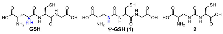
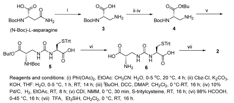
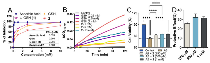
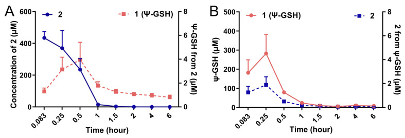
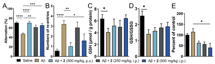
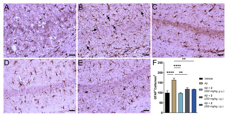
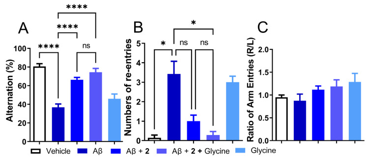
Supplementation of glutathione (GSH) levels through varying formulations or precursors has thus far appeared to be a tenable strategy to ameliorate disease-associated oxidative stress. Metabolic liability of GSH and its precursors, i.e., hydrolysis by the ubiquitous γ-glutamyl transpeptidase (γ-GT), has limited successful clinical translation due to poor bioavailability. We addressed this problem through the design of γ-GT-resistant GSH analogue, ψ-GSH, which successfully substituted in GSH-dependent enzymatic systems and also offered promise as a therapeutic for Alzheimer's disease (AD). With the aim to improve its bioavailability, we studied the utility of a ψ-GSH precursor, dipeptide 2, as a potential AD therapeutic. Compound 2 retains the γ-GT stable ureide linkage and the thiol group for antioxidant property. By engaging glutathione synthetase, compound 2 was able to generate ψ-GSH in vivo. It was found to be a modest cofactor of glutathione peroxidase and prevented cytotoxicity of Aβ1-42-aggregates in vitro. Studies of compound 2 in an acute AD model generated by intracerebroventricular injection of Aβ1-42 showed cognitive benefits, which were augmented by its combination with glycine along with mitigation of oxidative stress and inflammatory pathology. Collectively, these results support further optimization and evaluation of ψ-GSH dipeptide as a potential therapeutic in transgenic AD models.

Keywords: Alzheimer’s disease; antioxidant; glutathione; neuroinflammation; oxidative stress; precursor; ψ-GSH.


Conflict of interest statement

R.V., S.S.M., and A.R. are named inventors on the patent application relating to ψ-GSH and its analogs as treatment options of neurodegenerative disorders. The funders had no role in the design of the study; in the collection, analyses, or interpretation of data; in the writing of the manuscript, or in the decision to publish the results.

Figures

Figure 1
Figure 1
Chemical structures of ψ-GSH and the dipeptide of ψ-GSH.
Scheme 1
Scheme 1
Synthesis of ψ-GSH dipeptide (2).
Figure 2
Figure 2
Antioxidant property of the dipeptide of ψ-GSH (2) and its ability to protect against Aβ1–42 cytotoxicity in SH-SY5Y cells. (A) Analysis of antioxidant property of compound 2 by DPPH assay. (B) Glutathione peroxidase enzymatic assay. Measurement of glutathione peroxidase activity was performed using the purified enzyme in the presence of GSH or 2 as cofactors. In this coupled enzymatic assay, consumption of NADPH by glutathione reductase to reduce oxidized glutathione or 2 was measured. Differences in the levels of NADPH measured as absorbance at 340 nm compared to time zero are plotted. (C) Cytotoxicity of Aβ1–42 in SH-SY5Y cells was mitigated by co-incubation with compound 2 in a dose-dependent manner. Plot (D) shows dose-dependent protective effect of 2 calculated in relation to Aβ-only group. Data are presented as the mean ± SEM of three independent experiments. Treatment groups were analyzed for statistical significance using one-way ANOVA with Tukey’s post-hoc test. **** p < 0.0001.
Figure 3
Figure 3
Conversion of compound 2 to the tripeptide ψ-GSH (1) after administration in mice. Compounds 1 and 2 (250 mg/kg) were injected in 8–10-week old CD1 mice by intraperitoneal route. Quantitation of ψ-GSH (1) formed after injection of 2 (A) and vice versa (B) was performed using LC-MS/MS analysis. Formation of tripeptide 1 from the dipeptide 2 was detected, although at much lower levels.
Figure 4
Figure 4
Treatment with ψ-GSH dipeptide (2) restored cognitive impairment induced by i.c.v. Aβ1–42. After initiation of compound treatment, i.c.v. injection of Aβ1–42 was performed on day 4 and followed by continued treatment with compound 2 for 8 additional days. The T-maze spontaneous alternation test was conducted on days 10 and 11 to assess cognitive function. (A) Reduced alternation behavior was observed in saline treated Aβ-injected mice, which was restored to levels comparable to vehicle control mice with oral and i.p. treatment of 2. (B) Higher number of re-entries was evident in Aβ1–42-injected mice. Treatment with 2 (oral and high dose i.p.) showed reduction in repetitive arm entries. (C,D) Measurement of oxidative stress in the brain tissues was performed by analysis of reduced GSH and GSH/GSSG ratio. Compound-treated mice did not differ significantly from the vehicle-treated mice. (E) Quantitation of protein carbonyls by DNPH assay showed significant reduction in protein oxidation in the compound treated groups. Data are shown as the mean ± SEM. Statistical significance was assessed by a one-way ANOVA with Tukey’s post-hoc test (* p < 0.05, ** p < 0.01, *** p < 0.005, **** p < 0.001).
Figure 5
Figure 5
ψ-GSH dipeptide (2) treatment reduced reactive astrogliosis in i.c.v. Aβ1–42 injected mice. (AE) Representative images of reactive astrocytes visualized by GFAP antibody in hippocampus region of various treatment groups: saline (A), Aβ-only (B), Aβ + 2 (500 mg/kg, p.o., C), Aβ + 2 (250 mg/kg, i.p., D) Aβ + 2 (500 mg/kg, i.p., E). (F) Quantification of GFAP staining in the hippocampal region of all treatment groups. Data are shown as mean ± SEM. For comparisons between groups, a one-way ANOVA with Tukey’s post-hoc test was used for data analysis. ** p < 0.01, **** p < 0.001. Black arrows in panel B display typical GFAP positive staining of reactive astrocytes. All scale bars represent 25 μm.
Figure 6
Figure 6
Co-treatment with ψ-GSH dipeptide (2, 250 mg/kg, i.p.) with glycine (250 mg/kg, i.p.) showed improved cognitive benefits over the treatment of compound 2 alone. (A) Impaired alternation in Aβ-injected mice was restored by compound 2 treatment and showed a trend toward further improvement in the co-treatment (2 + glycine) group. (B) Increased number of re-entries in the Aβ-only treated group was significantly reduced in the co-treatment of compound 2 + glycine group, compared to treatment of compound 2 alone. (C) The treatment group did not show any side preference during the conduction of this test. Data are shown as the mean ± SEM. Statistical significance was assessed by a one-way ANOVA with Tukey’s post-hoc test (ns = not significant, * p < 0.05, **** p < 0.001).

References

    1. Tamagno E., Guglielmotto M., Vasciaveo V., Tabaton M. Oxidative stress and beta amyloid in alzheimer’s disease. Which comes first: The chicken or the egg? Antioxidants. 2021;10:1479. doi: 10.3390/antiox10091479. - DOI - PMC - PubMed
    1. Agostinho P., Cunha R.A., Oliveira C. Neuroinflammation, oxidative stress and the pathogenesis of alzheimer’s disease. Curr. Pharm. Des. 2010;16:2766–2778. doi: 10.2174/138161210793176572. - DOI - PubMed
    1. Aoyama K., Nakaki T. Impaired glutathione synthesis in neurodegeneration. Int. J. Mol. Sci. 2013;14:21021–21044. doi: 10.3390/ijms141021021. - DOI - PMC - PubMed
    1. Montine T.J., Neely M.D., Quinn J.F., Beal M.F., Markesbery W.R., Roberts L.J., Morrow J.D. Lipid peroxidation in aging brain and alzheimer’s disease. Free Radic. Biol. Med. 2002;33:620–626. - PubMed
    1. Markesbery W.R., Lovell M.A. Damage to lipids, proteins, DNA, and rna in mild cognitive impairment. Arch. Neurol. 2007;64:954–956. doi: 10.1001/archneur.64.7.954. - DOI - PubMed

LinkOut - more resources