Review
. 2022 Jun 13;11(6):1153.
doi: 10.3390/antiox11061153.

The Role of Reactive Oxygen Species in the Rheumatoid Arthritis-Associated Synovial Microenvironment

Affiliations
Review

The Role of Reactive Oxygen Species in the Rheumatoid Arthritis-Associated Synovial Microenvironment

Xing Wang et al. Antioxidants (Basel). .

Abstract

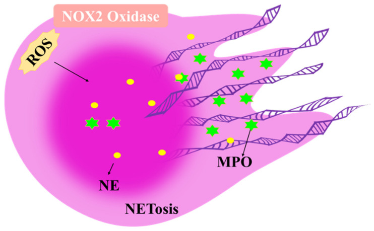
Rheumatoid arthritis (RA) is an inflammatory disease that begins with a loss of tolerance to modified self-antigens and immune system abnormalities, eventually leading to synovitis and bone and cartilage degradation. Reactive oxygen species (ROS) are commonly used as destructive or modifying agents of cellular components or they act as signaling molecules in the immune system. During the development of RA, a hypoxic and inflammatory situation in the synovium maintains ROS generation, which can be sustained by increased DNA damage and malfunctioning mitochondria in a feedback loop. Oxidative stress caused by abundant ROS production has also been shown to be associated with synovitis in RA. The goal of this review is to examine the functions of ROS and related molecular mechanisms in diverse cells in the synovial microenvironment of RA. The strategies relying on regulating ROS to treat RA are also reviewed.

Keywords: reactive oxygen species; rheumatoid arthritis; synovium; synovium microenvironment.


Conflict of interest statement

The authors declare no conflict of interest.

Figures

Figure 1
Figure 1
The synovial structure in healthy individuals and in individuals with rheumatoid arthritis (RA). (a) In the healthy joint, the synovial intimal lining consists of a thin cell layer of fibroblast synovial cells (FLSs) and macrophage-like synovial cells (MLSs) that together form a semi-permeable protective barrier. The sublining layer contains interstitial macrophages and fibroblasts, as well as blood vessels. (b) In RA, the macrophage barrier is lost and there is pathological expansion and remodeling of the synovial lining layer and sublining layer leading to synovial hyperplasia. The sublining of the synovium is heavily infiltrated by immune cells and undergoes neovascularization.
Figure 2
Figure 2
Role of reactive oxygen species (ROS)-induced gene mutations in fibroblast synovial cells. The synovial microenvironment of RA with hypoxia, high ROS and high inflammatory factors may lead to genetic imprinting of FLS by somatic and mitochondrial DNA mutations (as shown in the Red Cross), which lead to FLS proliferation and anti-apoptosis.
Figure 3
Figure 3
The role of ROS in macrophage-like synovial cells (MLSs). Under conditions, such as the inflammation and hypoxia that occur in RA joints, the MLSs switch to a high-load glycolytic metabolism, resulting in ROS leaking from the electron transport chain and the activation of pyruvate kinase M2 (PKM2), which in turn activates key downstream transcription factors, such as STAT3, NF-ΚB and hypoxia-inducible factor-1 α (HIF-1α), to drive TNF-α, IL-1β and IL-6 production.
Figure 4
Figure 4
The role of ROS in endothelial cells (ECs). Vascular endothelial growth factor (VEGF) stimulates ROS production through RAC1-mediated activation of NOX, followed by the autophosphorylation of VEGFR2 and activation of downstream signaling pathways critical for endothelial cell migration and proliferation. In addition, hypoxia activates the transcription factor HIF-1α by inducing ROS production, which in turn upregulates VEGF secretion and expression and promotes angiogenesis.
Figure 5
Figure 5
The role of ROS in neutrophils. ROS trigger the translocation of neutrophil elastase (NE) to the nucleus, where myeloperoxidase (MPO) then binds to chromatin and synergistically enhances chromatin deconcentration, leading to cell rupture and NET release.
Figure 6
Figure 6
The role of ROS in T cells. In T cells, inadequate activation of the DNA repair kinase ATM bypasses the G2/M cell cycle checkpoint due to intracellular ROS consumption. In addition, MRE11A inhibition leads to reduced mitochondrial ATP and ROS production, mtDNA deposition, caspase-1 activation and invasive tissue inflammation.

Similar articles

Cited by

References

    1. Klareskog L., Catrina A.I., Paget S. Rheumatoid arthritis. Lancet. 2009;373:659–672. doi: 10.1016/S0140-6736(09)60008-8. - DOI - PubMed
    1. Arnett F.C., Edworthy S.M., Bloch D.A., McShane D.J., Fries J.F., Cooper N.S., Healey L.A., Kaplan S.R., Liang M.H., Luthra H.S. The American Rheumatism Association 1987 revised criteria for the classification of rheumatoid arthritis. Arthritis Rheum. 1988;31:315–324. doi: 10.1002/art.1780310302. - DOI - PubMed
    1. Holers V.M., Demoruelle M.K., Kuhn K.A., Buckner J.H., Robinson W.H., Okamoto Y., Norris J.M., Deane K.D. Rheumatoid arthritis and the mucosal origins hypothesis: Protection turns to destruction. Nat. Rev. Rheumatol. 2018;14:542–557. doi: 10.1038/s41584-018-0070-0. - DOI - PMC - PubMed
    1. Buckley C.D., Ospelt C., Gay S., Midwood K.S. Location, location, location: How the tissue microenvironment affects inflammation in RA. Nat. Rev. Rheumatol. 2021;17:195–212. doi: 10.1038/s41584-020-00570-2. - DOI - PubMed
    1. O’Neil L.J., Kaplan M.J. Neutrophils in Rheumatoid Arthritis: Breaking Immune Tolerance and Fueling Disease. Trends Mol. Med. 2019;25:215–227. doi: 10.1016/j.molmed.2018.12.008. - DOI - PubMed

LinkOut - more resources