. 2022 Jun 14;11(6):1171.
doi: 10.3390/antiox11061171.

HO-1 Limits the Efficacy of Vemurafenib/PLX4032 in BRAFV600E Mutated Melanoma Cells Adapted to Physiological Normoxia or Hypoxia

Affiliations

HO-1 Limits the Efficacy of Vemurafenib/PLX4032 in BRAFV600E Mutated Melanoma Cells Adapted to Physiological Normoxia or Hypoxia

Anna Lisa Furfaro et al. Antioxidants (Basel). .

Abstract

Induction of heme oxygenase 1 (HO-1) favors immune-escape in BRAFV600 melanoma cells treated with Vemurafenib/PLX4032 under standard cell culture conditions. However, the oxygen tension under standard culture conditions (~18 kPa O2) is significantly higher than the physiological oxygen levels encountered in vivo. In addition, cancer cells in vivo are often modified by hypoxia. In this study, MeOV-1 primary melanoma cells bearing the BRAFV600E mutation, were adapted to either 5 kPa O2 (physiological normoxia) or 1 kPa O2 (hypoxia) and then exposed to 10 μM PLX4032. PLX4032 abolished ERK phosphorylation, reduced Bach1 expression and increased HO-1 levels independent of pericellular O2 tension. Moreover, cell viability was significantly reduced further in cells exposed to PLX4032 plus Tin mesoporphyrin IX, a HO-1 inhibitor. Notably, our findings provide the first evidence that HO-1 inhibition in combination with PLX4032 under physiological oxygen tension and hypoxia restores and increases the expression of the NK ligands ULBP3 and B7H6 compared to cells exposed to PLX4032 alone. Interestingly, although silencing NRF2 prevented PLX4032 induction of HO-1, other NRF2 targeted genes were unaffected, highlighting a pivotal role of HO-1 in melanoma resistance and immune escape. The present findings may enhance translation and highlight the potential of the HO-1 inhibitors in the therapy of BRAFV600 melanomas.

Keywords: HO-1; NK ligands; NRF2; hypoxia; melanoma; oxygen tension; physiological normoxia; response and/or resistance to therapy; target therapy.


Conflict of interest statement

The authors declare no conflict of interest.

Figures
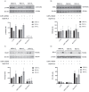
Figure 1
Figure 1
Immunoblot analysis of pERK (A), total ERK (B), Bach1 (C) and HO-1 (D). MeOV-1 cells were adapted for 5 days to 18 kPa, 5 kPa or 1 kPa O2 and then treated with 10 µM PLX4032 (or 0.05% DMSO) for 24 h. β-actin was used as loading control. Immunoblots are representative of 3–4 independent experiments. Data denote mean ± S.E.M., n = 3–4 independent cell cultures, § p < 0.001 vs. CTR; ★ p < 0.001 vs. DMSO; ★★ p < 0.01 vs. CTR; # p < 0.05 vs. DMSO; ## p < 0.01 vs. DMSO.
Figure 2
Figure 2
Melanoma cell viability after PLX4032 treatment and HO-1 inhibition. Cell viability was determined using the MTT assay in MeOV-1 cells seeded into 96-well plates and adapted for 5 days to 18 kPa O2, 5 kPa or 1 kPa O2. Cells were then treated with 10 µM PLX4032, 10 µM SnMP and combined treatment for 24 h. Data denote mean ± S.E.M., n = 5 independent cell cultures, ★ p < 0.005 vs. respective CTR and DMSO; # p < 0.05 vs. respective CTR and DMSO; § p < 0.05 vs. respective PLX4032; §§ p < 0.005 vs. respective PLX4032; §§§ p < 0.001 vs. respective PLX4032.
Figure 3
Figure 3
HIF-1α protein levels in MeOV-1 cells adapted for 5 days to different O2 levels. Immunoblot analysis of HIF-1α protein expression in MeOV-1 cells cultured for 5 days under 18 kPa O2, 5 kPa O2 or 1 kPa O2 and then treated with 10 µM PLX4032 (or 0.05% DMSO) and 10 µM SnMP for 24 h under the same O2 levels. β-actin was used as a loading control. Immunoblots are representative of 3 independent MeOV-1 cultures. Data denote mean ± S.E.M. ★ p < 0.05 vs. respective CTR and DMSO; § p < 0.005 vs. PLX under 18 kPa and PLX under 5 kPa O2.
Figure 4
Figure 4
mRNA expression of ULBP3 and B7H6 in MeOV-1 cells co-treated with PLX4032 and SnMP. ULBP3 (A) and B7H6 (B) mRNA expression was assessed by qPCR in MeOV-1 cells cultured under standard culture conditions (18 kPa O2) or adapted for 5 days to 5 kPa or 1 kPa O2. Cells were then exposed to 10 μM PLX4032 ± 10 μM SnMP for 24 h. Data shown are representative of 2 or 3 independent experiments for each experimental condition. Date denote mean ± S.E.M., ★ p < 0.05 vs. CTR and DMSO; # p < 0.05 vs. PLX4032.
Figure 5
Figure 5
PLX4032 induces NRF2-dependent HO-1 expression. HO-1, GCLC, NQO1 and GCLM mRNA expression was assessed by RT-PCR in MeOV-1 cells treated with 10 μM PLX4032 for 3 h and 6 h. Diethylmaleate (100 μM DEM) was used as a positive control and 18S mRNA was used as a housekeeping gene. Representative blots of 3 independent experiments are shown in (A). Densitometric analysis for each gene shown in (B) where data denote mean ± S.E.M. ★ p < 0.05 vs. respective CTR; ★★ p < 0.001 vs. CTR. The results of NRF2 silencing on HO-1 expression are shown in (C): HO-1 protein (upper) and mRNA (lower) expression were assessed by immunoblotting and by RT-PCR, using DEM as a positive control. Actin protein level and 18S mRNA were used as internal controls, respectively. Representative blots of 3 independent experiments are shown, with densitometric analyses of HO-1 mRNA expression. Data denote mean ± S.E.M. s p < 0.005 vs. PLX.

Similar articles

Cited by

References

    1. Marengo B., Nitti M., Furfaro A.L., Colla R., Ciucis C.D., Marinari U.M., Pronzato M.A., Traverso N., Domenicotti C. Redox Homeostasis and Cellular Antioxidant Systems: Crucial Players in Cancer Growth and Therapy. Oxid. Med. Cell. Longev. 2016;2016:6235641. doi: 10.1155/2016/6235641. - DOI - PMC - PubMed
    1. Helfinger V., Schröder K. Redox Control in Cancer Development and Progression. Mol. Asp. Med. 2018;63:88–98. doi: 10.1016/j.mam.2018.02.003. - DOI - PubMed
    1. Jozkowicz A., Was H., Dulak J. Heme Oxygenase-1 in Tumors: Is It a False Friend? Antioxid. Redox Signal. 2007;9:2099–2117. doi: 10.1089/ars.2007.1659. - DOI - PMC - PubMed
    1. Traverso N., Ricciarelli R., Nitti M., Marengo B., Furfaro A.L., Pronzato M.A., Marinari U.M., Domenicotti C. Role of Glutathione in Cancer Progression and Chemoresistance. Oxid. Med. Cell. Longev. 2013;2013:972913. doi: 10.1155/2013/972913. - DOI - PMC - PubMed
    1. Nitti M., Piras S., Marinari U.M., Moretta L., Pronzato M.A., Furfaro A.L. HO-1 Induction in Cancer Progression: A Matter of Cell Adaptation. Antioxidants. 2017;6:29. doi: 10.3390/antiox6020029. - DOI - PMC - PubMed