. 2022 Jun 15;11(6):1177.
doi: 10.3390/antiox11061177.

Exogenous Melatonin in the Culture Medium Does Not Affect the Development of In Vivo-Derived Pig Embryos but Substantially Improves the Quality of In Vitro-Produced Embryos

Affiliations

Exogenous Melatonin in the Culture Medium Does Not Affect the Development of In Vivo-Derived Pig Embryos but Substantially Improves the Quality of In Vitro-Produced Embryos

Cristina A Martinez et al. Antioxidants (Basel). .

Abstract

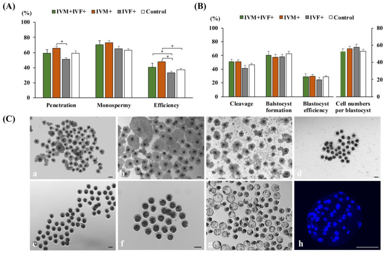
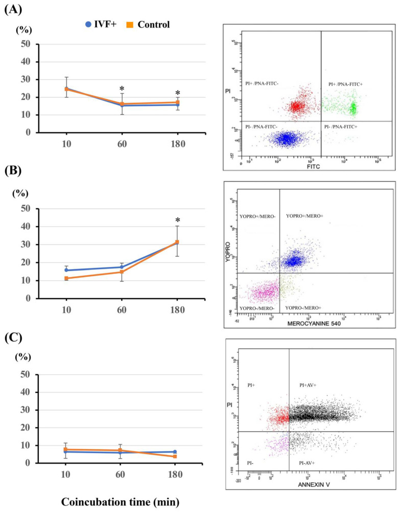
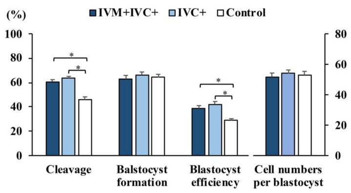
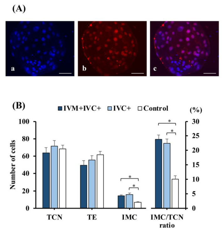
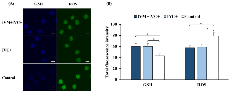
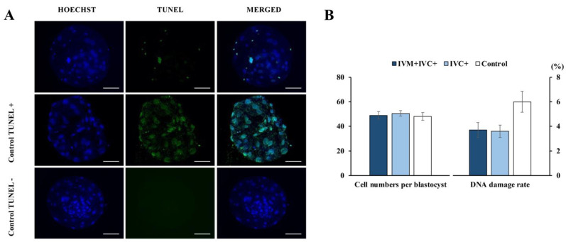
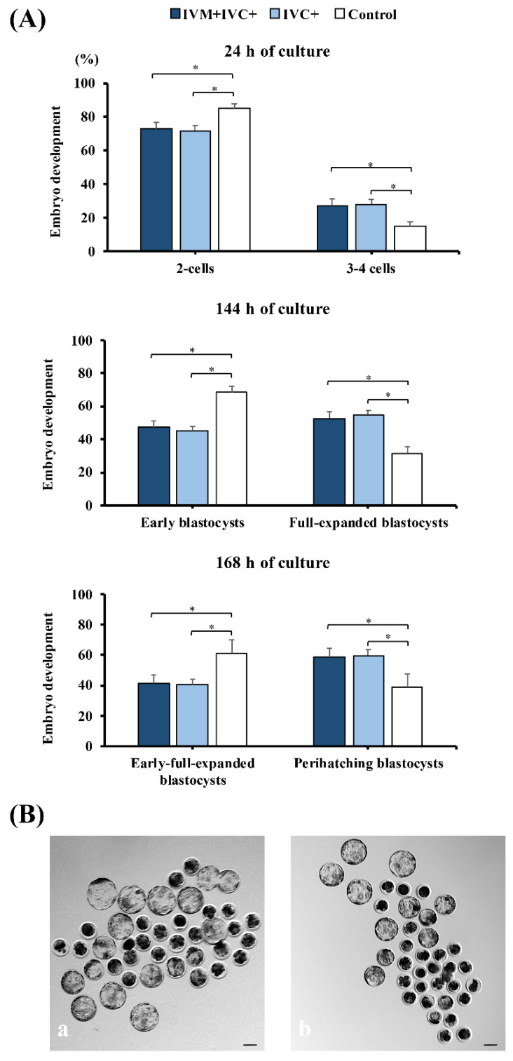
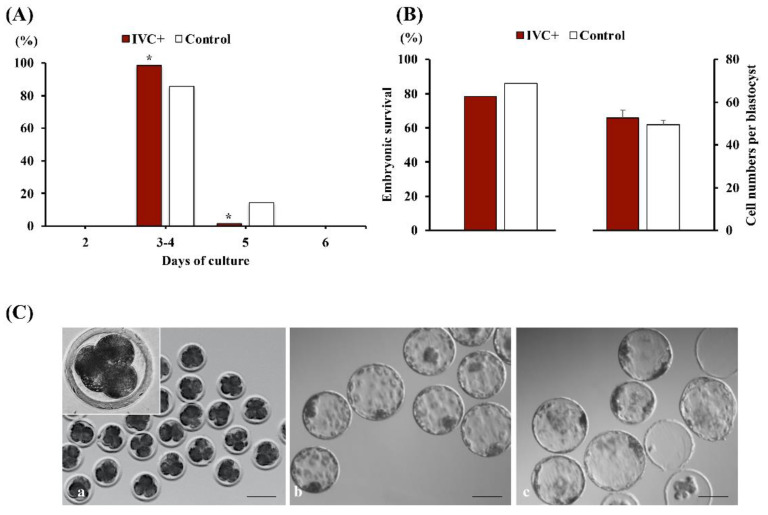
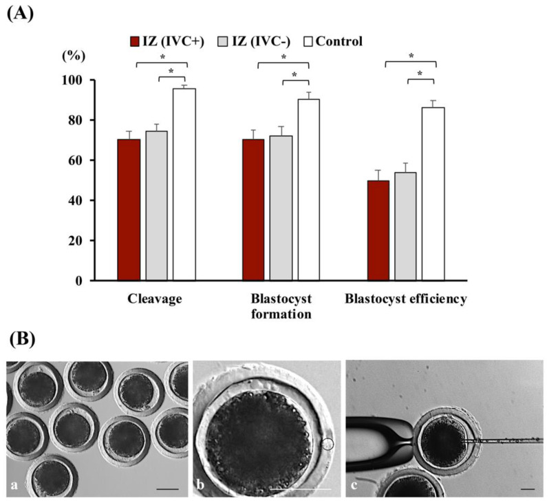
Cloned and transgenic pigs are relevant human disease models and serve as potential donors for regenerative medicine and xenotransplantation. These technologies demand oocytes and embryos of good quality. However, the current protocols for in vitro production (IVP) of pig embryos give reduced blastocyst efficiency and embryo quality compared to in vivo controls. This is likely due to culture conditions jeopardizing embryonic homeostasis including the effect of reactive oxygen species (ROS) influence. In this study, the antioxidant melatonin (1 nM) in the maturation medium, fertilization medium, or both media was ineffective in enhancing fertilization or embryonic development parameters of in vitro fertilized oocytes. Supplementation of melatonin in the fertilization medium also had no effect on sperm function. In contrast, the addition of melatonin to the embryo culture medium accelerated the timing of embryonic development and increased the percentages of cleaved embryos and presumed zygotes that developed to the blastocyst stage. Furthermore, it increased the number of inner mass cells and the inner mass cell/total cell number ratio per blastocyst while increasing intracellular glutathione and reducing ROS and DNA damage levels in embryos. Contrarily, the addition of melatonin to the embryo culture medium had no evident effect on in vivo-derived embryos, including the developmental capacity and the quality of in vivo-derived 4-cell embryos or the percentage of genome-edited in vivo-derived zygotes achieving the blastocyst stage. In conclusion, exogenous melatonin in the embryo culture medium enhances the development and quality of in vitro-derived embryos but not in in vivo-derived embryos. Exogenous melatonin is thus recommended during embryo culture of oocytes matured and fertilized in vitro for improving porcine IVP efficiency.

Keywords: CRISPR/Cas9; apoptosis; in vitro fertilization; in vivo-derived embryos; inner cell mass; melatonin; pig; reactive oxygen species.


Conflict of interest statement

The authors declare no conflict of interest. The funders had no role in the design of the study; in the collection, analyses, or interpretation of data; in the writing of the manuscript, or in the decision to publish the results.

Figures

Figure 1
Figure 1
Effects of melatonin (1 nM) supplementation of in vitro maturation (IVM) and/or in vitro fertilization (IVF) medium on the development of porcine oocytes. (A) Fertilization parameters. IVM + IVC+: melatonin added to both IVM and IVF media (n = 132); IVM+: melatonin only added to IVM medium (n = 131); IVF+: melatonin only added to the IVF medium (n = 135); control: untreated (n = 134). Penetration: number of penetrated oocytes relative to the number of mature oocytes inseminated; monospermy: number of monospermic oocytes relative to the number of mature oocytes penetrated; efficiency: number of monospermic oocytes relative to the number of mature oocytes inseminated. Data are presented as the means ± SEM of six replicates. * p < 0.025. (B) Embryonic development parameters. IVM + IVC+ (n = 249); IVM+ (n = 247); IVF+ (n = 250); control (n = 258). The number of blastocysts evaluated to determine the total cell number in each group was 70, 76, 60, and 73, respectively. Cleavage: percentage of oocytes inseminated that developed to the 2- to 4-cell embryos at 48 h of culture; Blastocyst formation: percentage of cleaved embryos that progressed to blastocysts at day 7 of culture; blastocyst efficiency: percentage of oocytes inseminated that progressed to blastocyst. Data are presented as the means ± SEM of six replicates. (C) Representative images of immature oocytes (a), oocytes at 22 h and 44 h of maturation ((b,c) respectively), oocytes during the coincubation period (d), presumed zygotes after IVF (e), cleaved embryos, and blastocyst development at days 2 and 7 of culture ((f,g) respectively) and Hoechst-stained nuclei of an in vitro-produced blastocyst (h). Scale bar: 100 µm.
Figure 2
Figure 2
Effects of 1 nM melatonin addition to gamete coincubation medium (IVF+) on sperm parameters. (A) Sperm viability: viable sperm population exhibiting intact plasma and acrosomal membranes (PI−/PNA-FITC−); (B) Plasma membrane destabilization: viable sperm population with high fluidity of the plasma membrane (YOPRO−/MERO+); (C) Apoptosis: viable spermatozoa displaying early apoptotic-like changes (PI−/AV+). Gametes coincubated in the absence of melatonin constituted the control group. * Different from the first value (p < 0.05). Data are presented as the means ± SEM of three replicates.
Figure 3
Figure 3
Effects of melatonin (1 nM) supplementation of in vitro maturation (IVM) and/or in vitro embryo culture (IVC) medium on the development of porcine zygotes. IVM + IVC+: melatonin added to both IVM and IVC media (n = 410); IVC+: melatonin added only to the IVC medium (n = 406); control: untreated (n = 409). The number of blastocysts evaluated to determine the total cell number in each group was 97, 105, and 93, respectively. Cleavage: percentage of oocytes inseminated that developed to the 2- to 4-cell embryos at 48 h of culture; blastocyst formation: percentage of cleaved embryos that progressed to blastocyst at day 7 of culture; blastocyst efficiency: percentage of oocytes inseminated that progressed to blastocysts. * p < 0.006. Data are presented as the means ± SEM of six replicates.
Figure 4
Figure 4
Differential staining of day 7 blastocysts produced in vitro. (A) Fluorescence microscopy image showing a blastocyst subjected to differential staining to determine the total cell number (TCN) and the number of trophectoderm (TE) cells, which were stained with Hoechst 33,342 (blue nuclei); (a) and an anti-CDX2 antibody (red nuclei); (b) Merged images (c) show the inner cell mass (ICM) and TE cells with blue and pink-red fluorescence, respectively. Scale bar: 50 µm. (B) Distribution of the different types of cells in blastocysts produced by adding melatonin (1 nM) to both the maturation and culture media (IVM + IVC+ group; n = 14), or only to the culture medium (IVC+ group; n = 15). Blastocysts derived from oocytes that were matured, fertilized, and cultured in media without melatonin supplementation were used as controls (n = 15). * p < 0.001. Data are presented as the means ± SEM (three replicates).
Figure 5
Figure 5
Effects of melatonin on intracellular glutathione (GSH) and reactive oxygen species (ROS) levels in in vitro-produced 4-cell embryos at 48 h of culture. (A) Representative images of embryos stained with CellTracker Blue (GSH) or with 2′,7′-dichlorodihydrofluorescein diacetate (ROS). Scale bar: 100 µm. (B) Effects of melatonin on GSH and ROS levels in embryos. Melatonin (1 nM) was added to both the maturation and culture media (IVM + IVC+ group; n = 28) or only to the culture medium (IVC+ group; n = 32). Oocytes that were matured, fertilized, and cultured in media without melatonin supplementation were used as controls (n = 30). * p < 0.03. Data are presented as the means ± SEM (three replicates).
Figure 6
Figure 6
Detection of DNA damage in in vitro-produced Day 7 blastocysts. (A) Representative fluorescence images of the TUNEL assay. Hoechst (blue) and TUNEL (green) staining indicate the total cells and the cells with DNA damage, respectively. Scale bar: 50 µm. (B) Effects of melatonin on DNA damage rates in blastocysts. Melatonin (1 nM) was added to both the maturation and culture media (IVM + IVC+ group; n = 31) or only to the culture medium (IVC+ group; n = 30). Oocytes matured, fertilized, and cultured in media without melatonin supplementation were used as controls (n = 26). Data are expressed as the mean ± SEM (three replicates).
Figure 7
Figure 7
Embryonic developmental stages at 24 h, 144 h, and 168 h of culture were achieved by oocytes and embryos exposed or not exposed (control) to 1 nM melatonin. (A) Percentages of 3, 4-cell embryos at 24 h of culture from the total number of cleaved embryos at 48 h of culture and distribution of the different blastocyst stages reached in each group at 144 h and 168 h of culture, respectively. IVM + IVC+ group (n = 323): maturation and embryo culture performed in media supplemented with melatonin; IVC+ group (n = 310): embryo culture performed in medium containing melatonin. Control group (n = 295): maturation and embryo culture performed without melatonin. Perihatching blastocysts include hatching and hatched blastocysts. * p < 0.05. Data are presented as the means ± SEM (four replicates). (B) Representative images of blastocyst development at 168 h of culture in the presence (a) or absence (b) of melatonin. Scale bar: 100 µm.
Figure 8
Figure 8
Effect of melatonin on the in vitro development of in vivo-derived 4-cell embryos. (A) Effects of the addition of 1 nM melatonin to the culture medium on the percentages of in vivo-derived 4-cell embryos developing to the expanded blastocyst stage or later stages at different days of culture. The embryos were cultured in culture medium with (IVC+; n = 70) or without (control; n = 76) melatonin. * p < 0.004 compared to the control group. (B) Post-warming survival and total cell number per blastocyst of vitrified-warmed expanded blastocysts formed from in vivo-derived 4-cell embryos cultured in a medium supplemented with (IVC+; n = 68) or without (control; n = 76) melatonin. Data are presented as percentages and means ± SEM (four replicates). (C) Representative images of in vivo-derived 4-cell embryos (a), fresh (b), and vitrified-warmed (c) blastocysts produced from the in vitro culture of in vivo-derived 4-cell embryos. Scale bar: 100 µm.
Figure 9
Figure 9
Effect of melatonin on the in vitro development of microinjected zygotes produced in vivo. (A) Cleavage, blastocyst formation, and blastocyst efficiency rates of in vivo-derived, injected zygotes (IZ) cultured for 168 h in a culture medium containing (IVC+; n = 217) or lacking (IVC−; n = 214) 1 nM melatonin. Noninjected in vivo-derived zygotes cultured in the absence of melatonin were used as controls (n = 65). * p < 0.001. Data are presented as the means ± SEM of six replicates. (B) Representative images of in vivo-derived zygotes (a) and microinjection procedure (b,c). Scale bar: 50 µm.

Similar articles

Cited by

References

    1. Gil M.A., Cuello C., Parrilla I., Vazquez J.M., Roca J., Martinez E.A. Advances in Swine In Vitro Embryo Production Technologies. Reprod. Domest. Anim. 2010;45:40–48. doi: 10.1111/j.1439-0531.2010.01623.x. - DOI - PubMed
    1. Grupen C.G. The evolution of porcine embryo in vitro production. Theriogenology. 2014;81:24–37. doi: 10.1016/j.theriogenology.2013.09.022. - DOI - PubMed
    1. Fowler K.E., Mandawala A.A., Griffin D.K., Walling G.A., Harvey S.C. The production of pig preimplantation embryos in vitro: Current progress and future prospects. Reprod. Biol. 2018;18:203–211. doi: 10.1016/j.repbio.2018.07.001. - DOI - PubMed
    1. Martinez C.A., Martinez E.A., Gil M.A. Importance of oil overlay for production of porcine embryos in vitro. Reprod. Domest. Anim. 2018;53:281–286. doi: 10.1111/rda.13114. - DOI - PubMed
    1. Macháty Z., Day B.N., Prather R.S. Development of early porcine embryos in vitro and in vivo. Biol. Reprod. 1998;59:451–455. doi: 10.1095/biolreprod59.2.451. - DOI - PubMed