. 2022 Jun 16;11(6):1186.
doi: 10.3390/antiox11061186.

Preclinical Characterization of Antioxidant Quinolyl Nitrone QN23 as a New Candidate for the Treatment of Ischemic Stroke

Affiliations

Preclinical Characterization of Antioxidant Quinolyl Nitrone QN23 as a New Candidate for the Treatment of Ischemic Stroke

Emma Martínez-Alonso et al. Antioxidants (Basel). .

Abstract

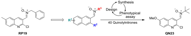
Nitrones are encouraging drug candidates for the treatment of oxidative stress-driven diseases such as acute ischemic stroke (AIS). In a previous study, we found a promising quinolylnitrone, QN23, which exerted a neuroprotective effect in neuronal cell cultures subjected to oxygen-glucose deprivation and in experimental models of cerebral ischemia. In this paper, we update the biological and pharmacological characterization of QN23. We describe the suitability of intravenous administration of QN23 to induce neuroprotection in transitory four-vessel occlusion (4VO) and middle cerebral artery occlusion (tMCAO) experimental models of brain ischemia by assessing neuronal death, apoptosis induction, and infarct area, as well as neurofunctional outcomes. QN23 significantly decreased the neuronal death and apoptosis induced by the ischemic episode in a dose-dependent manner and showed a therapeutic effect when administered up to 3 h after post-ischemic reperfusion onset, effects that remained 11 weeks after the ischemic episode. In addition, QN23 significantly reduced infarct volume, thus recovering the motor function in a tMCAO model. Remarkably, we assessed the antioxidant activity of QN23 in vivo using dihydroethidium as a molecular probe for radical species. Finally, we describe QN23 pharmacokinetic parameters. All these results pointing to QN23 as an interesting and promising preclinical candidate for the treatment of AIS.

Keywords: antioxidants; brain ischemia; ischemic stroke; neuroprotection; pharmacokinetics; quinolyl nitrones; reactive oxygen species.


Conflict of interest statement

The authors declare no conflict of interest. The funder Isquaemia Biotech SL did not have role in the study design, data collection and analysis or interpretation, in the preparation of the manuscript, or in the decision to publish the results.

Figures

Scheme 1
Scheme 1
Development of QN23.
Figure 1
Figure 1
Dose–response curve of QN23. Ischemic animals were treated with vehicle or QN23 (1.0–2.5 mg/kg) by intravenous injection at reperfusion onset after global cerebral ischemia, and the effect of QN23 was studied by assessing neuronal death and apoptosis after 5 days of reperfusion (R5d). (a) Representative images of Fluoro-Jade B-stained brain sections from vehicle- or QN23-treated animals (R5d + vehicle and R5d + QN23, respectively). Fluoro-Jade B-positive dead neurons were visualized by fluorescence microscopy (in green) in the hippocampal CA1 (CA1) and cortical (C) regions, and counted as described in the Materials and Methods section. (b) Bar graph representation of the neuronal death quantified in brain sections stained with Fluoro-Jade B in CA1 and C regions. Data represent the mean of 9 independent animals treated with vehicle, and 6–7 independent animals per QN23 treatment group; error bars indicate the SE. * p < 0.05 and ** p < 0.01, compared with the R5d + vehicle (Vh) by Dunnett’s post-test after ANOVA. In (c), data from (b) are represented as neuroprotective activity of QN23. Neuroprotection was defined as the effect achieved relative to the neuronal death value observed in the R5d + vehicle group (Vh) (0% value) and to the absence of neuronal death (100% value). Data are represented as mean ± SE. (d) Brain sections as in (a) were used for apoptosis detection by TUNEL assay and visualized by fluorescence microscopy (in green) in CA1 and C regions. Images are representative of the TUNEL assay, with the corresponding Hoechst counterstaining. TUNEL-positive cells were counted in CA1 and C regions as described in the Materials and Methods section. (e) Bar graph representation of neuronal apoptosis quantification, performed as in (b). In (f), data from (e) are represented as in (c).
Figure 2
Figure 2
Neurodeficit score outcomes after cerebral ischemia in the dose–response study of QN23. Bar graph representation of the neurological deficit score (NDS) in ischemic animals at 5 days of reperfusion after ischemia. Animals were treated with vehicle (Vh) or QN23 in 1.0, 1.5, 2.0, or 2.5 mg/kg by an intravenous injection at reperfusion onset after cerebral ischemia. A total of 6–9 independent animals (as described in Figure 1) were averaged per group; error bars indicate the SE, * p < 0.05, compared with vehicle by Dunn’s post-test after the non-parametric Kruskal–Wallis test.
Figure 3
Figure 3
Antioxidant capacity of QN23 in vivo. Ischemic animals were treated with QN23 (2.0 mg/kg) or vehicle (Vh) by intravenous injection at the onset of reperfusion after cerebral ischemia (IR), dihydroethidium (DHE, 3.0 mg/kg) was injected intravenously at 2 h after reperfusion, and the animals were euthanized at 4 h after DHE injection. Control animals were also injected with DHE and euthanized 4 h after injection. The antioxidant effect of QN23 was studied by quantification of fluorescence intensity of ROS-reacted DHE in brain sections. Representative images of cells stained with ROS-reacted DHE (DHE+ cells) in the hippocampal CA1 (a) and cortical (b) regions. Bar graphs represent the fluorescence intensity (FI) quantified in brain sections from control, vehicle-, and QN23-treated animals injected with DHE in the hippocampal CA1 (CA1) and cortical (C) regions. Data represent the mean of 3–5 independent animals per group (3 control animals, 5 animals treated with vehicle, and 4 treated with QN23); error bars indicate the SE. * p < 0.05 and ** p < 0.01, compared with the control or QN23-treated group by Newman–Keuls’ post-test after ANOVA. FI is expressed in arbitrary units (a.u.).
Figure 4
Figure 4
Therapeutic window of QN23 studied by Fluoro-Jade B assay (neuronal death). Ischemic animals were treated with vehicle (Vh), QN23 (2.0 mg/kg), or NXY-059 (40 mg/kg) by an intravenous injection at reperfusion onset (0 h), or at 1, 3, or 6 h of reperfusion after global cerebral ischemia. Neuroprotection was evaluated after 5 days of post-ischemic reperfusion. (a) Representative images of Fluoro-Jade B-stained hippocampal CA1 (CA1) and cortical (C) brain sections of vehicle- and QN23-treated animals at 1 h or 3 h of reperfusion and NXY-059-treated animals at 0 h of reperfusion. Dead neurons observed after Fluoro-Jade B staining within the CA1 and C regions were imaged by fluorescence microscopy (in green) and counted as described in the Materials and Methods section. (b) Bar graphs represent the neuronal death quantified in brain sections from vehicle-, QN23-, and NXY-059-treated animals in CA1 and C regions. For each group, 6–10 independent animals were averaged (10 animals treated with vehicle, 6–7 animals per QN23 treatment, and 6 animals per NXY-059 treatment); error bars indicate the SE. * p < 0.05 and ** p < 0.01, compared with vehicle (Vh) by Dunnett’s post-test, and # p < 0.05, by Student’s t test, after ANOVA.
Figure 5
Figure 5
Therapeutic window of QN23 studied by TUNEL assay (apoptosis). Brain sections of ischemic animals from Figure 4 were used for apoptosis detection by TUNEL assay in the hippocampal CA1 (CA1) and cortical (C) regions and imaged by fluorescence microscopy (in green). (a) Representative images of CA1 or C regions from vehicle- and QN23-treated animals at 1 h or 3 h of reperfusion and NXY-059-treated animals at 0 h of reperfusion, after apoptosis detection by TUNEL assay and the corresponding Hoechst counterstaining. TUNEL-positive cells detected within CA1 and C regions were counted as described in the Materials and Methods section. (b) Bar graph representations of TUNEL-positive cells quantified in brain sections from vehicle-, QN23-, and NXY-059-treated animals in CA1 and C regions. For each group, 6–10 independent animals (as described in Figure 4) were averaged; error bars indicate the SE. * p < 0.05 and ** p < 0.01, compared with vehicle (Vh) by Dunnett’s post-test, after ANOVA.
Figure 6
Figure 6
Neurodeficit score outcomes after cerebral ischemia in the therapeutic window study of QN23. The bar graph shows the neurological deficit score (NDS) in ischemic animals after 5 days of reperfusion after ischemia. Animals were treated with QN23 (2.0 mg/kg), NXY-059 (40 mg/kg), or vehicle (Vh) by intravenous injection at the reperfusion onset (0 h), or after 1, 3, or 6 h of post-ischemic reperfusion. For each group, 6–10 independent animals (as described in Figure 4) were averaged; error bars indicate the SE, * p < 0.05 and ** p < 0.01 compared with vehicle by Dunn’s post-test after Kruskal–Wallis test.
Figure 7
Figure 7
Study of the long-term effect of QN23 after cerebral ischemia–reperfusion by neuronal viability assessment. Brain sections from control animals (control) or from animals treated with vehicle (Vh), QN23 (2.0 mg/kg), or NXY-059 (40 mg/kg) by intravenous injection at the reperfusion onset after ischemia were evaluated for immunostaining at 11 weeks after ischemic reperfusion (IR). Neurons were labeled with anti-S6 protein antibody and X Red-secondary antibody and visualized by fluorescence microscopy (in red). Cell nuclei were counterstained with Hoechst dye (in blue). (a) Representative images of the hippocampal CA1 region. (b) Dot graph representation of S6-labeled cells (S6+ cells) per field of the hippocampal CA1 region from control animals and vehicle (Vh)-, QN23-, and NXY-059-treated animals after 11 weeks of post-ischemic reperfusion. Individual values of 6 independent animals per group are shown and the horizontal line represents the mean. ** p < 0.01, compared with the control group by Dunnett’s post-test after ANOVA.
Figure 8
Figure 8
Study of the long-term effect of QN23 after cerebral ischemia–reperfusion by behavioral tests. Graphic representations of the results of three tests assessing the spatial recognition and spatial memory, as long-term cognitive sequelae, of control, vehicle-, QN23-, and NXY-059-treated animals 11 weeks after post-ischemic reperfusion. In (a,b), individual values of 6 independent animals per group are shown and the horizontal line represents the mean. * p < 0.05, compared with the control group by Dunn’s post-test after the non-parametric Kruskal–Wallis test, *** p < 0.001, compared with the vehicle (Vh) group by Fisher’s test. In (c), two animals of the control group had a null test.
Figure 9
Figure 9
Cerebroprotective effects of QN23 in male Wistar rats subjected to ischemic stroke and treated with vehicle (Vh, n = 7); QN23, 1.5 mg/kg (n = 7); QN23, 2.5 mg/kg (n = 7); and QN23 4.0 mg/kg (n = 5). (a) Neurofunctional score at 24 h and 48 h after tMCAO (transient middle cerebral artery occlusion). (b) Representative images of 2 mm-thick coronal sections (0.2 to −1.8 mm from bregma) stained with 2,3,5-triphenyltetrazolium chloride (TTC) to show the infarcted (pale) area at 48 h after tMCAO. (c) Cortical, subcortical, and total infarct volumes at 48 h after tMCAO. (d) Edema volume at 48 h after tMCAO. Statistical significance compared with vehicle, * p < 0.05 and ** p < 0.01, was done between vehicle and 1.5 mg/kg and 2.5 mg/kg QN23-treated groups by Dunn’s post-test after the non-parametric Kruskal–Wallis test in (a), and by Dunnett’s post-test after ANOVA in (c,d).
Figure 10
Figure 10
QN23 plasma concentration–time profile after intravenous administration and values of AUC0–8h (AUCt) and Cmax versus dose. Data represented as mean ± SE of 6 independent animals. AUC, area under the curve.

References

    1. World Health Organization . Global Health Estimates 2020: Deaths by Cause, Age, Sex, by Country and by Region, 2000–2019. World Health Organization; Geneva, Switzerland: 2020.
    1. Moskowitz M.A., Lo E.H., Iadecola C. The science of stroke: Mechanisms in search of treatments. Neuron. 2010;67:181–198. doi: 10.1016/j.neuron.2010.07.002. - DOI - PMC - PubMed
    1. Powers W.J., Rabinstein A.A., Ackerson T., Adeoye O.M., Bambakidis N.C., Becker K., Biller J., Brown M., Demaerschalk B.M., Hoh B., et al. Guidelines for the Early Management of Patients with Acute Ischemic Stroke: 2019 Update to the 2018 Guidelines for the Early Management of Acute Ischemic Stroke: A Guideline for Healthcare Professionals From the American Heart Association/American Stroke Association. Stroke. 2019;50:e344–e418. doi: 10.1161/STR.0000000000000211. - DOI - PubMed
    1. White B.C., Sullivan J.M., DeGracia D.J., O’Neil B.J., Neumar R.W., Grossman L.I., Rafols J.A., Krause G.S. Brain ischemia and reperfusion: Molecular mechanisms of neuronal injury. J. Neurol. Sci. 2000;179:1–33. doi: 10.1016/S0022-510X(00)00386-5. - DOI - PubMed
    1. Brouns R., De Deyn P.P. The Complexity of Neurobiological Processes in Acute Ischemic Stroke. Clin. Neurol. Neurosurg. 2009;111:483–495. doi: 10.1016/j.clineuro.2009.04.001. - DOI - PubMed

LinkOut - more resources