. 2022 May 24;10(6):1217.
doi: 10.3390/biomedicines10061217.

Antitumor Properties of a New Macrocyclic Tetranuclear Oxidovanadium(V) Complex with 3-Methoxysalicylidenvaline Ligand

Affiliations

Antitumor Properties of a New Macrocyclic Tetranuclear Oxidovanadium(V) Complex with 3-Methoxysalicylidenvaline Ligand

Mihaela Turtoi et al. Biomedicines. .

Abstract

A wide variety of metal-based compounds have been obtained and studied for their antitumor activity since the intensely used cytostatic drugs (e.g., cisplatin) failed to accomplish their expected pharmacological properties. Thus, we aimed to develop a new vanadium-based drug and assess its antitumor properties using the human hepatocarcinoma (HepG2) cell line. The compound was synthesized from vanadyl sulfate, DL-valine, and o-vanillin and was spectrally and structurally characterized (UV-Vis, IR, CD, and single-crystal/powder-XRD). Compound stability in biological media, cell uptake, and the interaction with albumin were assessed. The mechanisms of its antitumor activity were determined compared to cisplatin by performing cytotoxicity, oxidative and mitochondrial status, DNA fragmentation, β-Tubulin synthesis investigation, and cell cycle studies. Herein, we developed a macrocyclic tetranuclear oxidovanadium(V) compound, [(VVO)(L)(CH3O)]4, having coordinated four Schiff base (H2L) ligands, 3-methoxysalicylidenvaline. We showed that [(VVO)(L)(CH3O)]4: (i) has pH-dependent stability in biological media, (ii) binds to albumin in a dose-dependent manner, (iii) is taken up by cells in a time-dependent way, (iv) has a higher capacity to induce cell death compared to cisplatin (IC50 = 6 μM vs. 10 μM), by altering the oxidative and mitochondrial status in HepG2 cells. Unlike cisplatin, which blocks the cell cycle in the S-phase, the new vanadium-based compound arrests it in S and G2/M-phase, whereas no differences in the induction of DNA fragmentation and reduction of β-Tubulin synthesis between the two were determined. Thus, the [(VVO)(L)(CH3O)]4 antitumor mechanism involved corroboration between the generation of oxidative species, mitochondrial dysfunction, degradation of DNA, cell cycle arrest in the S and G2/M-phase, and β-Tubulin synthesis reduction. Our studies demonstrate the potent antitumor activity of [(VVO)(L)(CH3O)]4 and propose it as an attractive candidate for anticancer therapy.

Keywords: 3-methoxysalicylidenvaline ligand; antitumor activity; apoptosis; hepatocarcinoma; oxidovanadium(V) complex.


Conflict of interest statement

The authors of this paper have nothing to disclose.

Figures

Figure 1
Figure 1
The X-ray crystal structure of TetraVV with the atom labeling scheme of non-carbon atoms. For clarity, hydrogen atoms have been excluded from the diagram.
Figure 2
Figure 2
The experimental and simulated powder X-ray diffractograms for compound TetraVV.
Figure 3
Figure 3
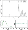
(A) IR spectrum of TetraVV. (B) Electronic spectrum of 100 μM TetraVV in phosphate-buffered solution (PBS), pH = 7.4. (C) CD spectrum of 500 μM TetraVV in PBS, pH = 7.4.
Figure 4
Figure 4
(A) Fluorescence spectra of 2 µM BSA in the presence of 0.1% DMSO or 1–25 µM TetraVV. (B) The electronic spectra of 100 μM TetraVV in PBS, pH = 7.4 vs. DMEM culture medium, pH = 7.4. (C) Time dependent stability of 100 μM TetraVV in DMEM medium at pH = 7.4 and pH = 4.5. (D) The concentration of TetraVV measured at 0, 4 and 24 h in DMEM at pH = 7.4 and pH = 4.5. Data were expressed as mean ± SD: ** p < 0.01, **** p < 0.0001.
Figure 5
Figure 5
(A) The cell viability of HepG2 cells treated with various concentrations (0.25–64 µM) of TetraVV and cisplatin. (B) Adenylate kinase (AK) release expressed as fold change to Control. (C) The number of dead cells (red fluorescence)/total cell no. (red + green fluorescence) calculated from Calcein-AM/propidium iodide (PI) staining and expressed as fold change to Control. (D) Representative images of calcein-AM (green)/PI (red) staining. Scale bar = 100 µm. HepG2 cells were treated with IC50 concentration of TetraVV and cisplatin (6 µM and 10 µM, respectively) or exposed to 0.01% DMSO (vehicle, Control) for 24 h. Data were expressed as mean ± SD: **** p < 0.0001 vs. Control, ## p < 0.01, #### p < 0.0001 vs. Cisplatin.
Figure 6
Figure 6
(A) Vanadium(V) content (ng) per 105 HepG2 cells over 24 h. (B) Adenylate kinase (AK) release expressed as fold change to Control. (C) The cellular morphology of HepG2 cells exposed to TetraVV and cisplatin for up to 24 h. Scale bar = 200 µm. HepG2 cells were treated with IC50 concentration of TetraVV or cisplatin (6 µM or 10 µM, respectively) or exposed to 0.01% DMSO (vehicle, Control) for 4 and 24 h. Data were expressed as mean ± SD: **** p < 0.0001 vs. 4 h.
Figure 7
Figure 7
The intracellular levels of ROS expressed as DCFH-DA fluorescence intensity/µg protein (relative to control cells) in HepG2 cells at 4 h (A) and 24 h (B). The extracellular levels of MDA (relative to control cells) in HepG2 cells at 4 h (C) and 24 h (D). HepG2 cells were treated with 6 µM TetraVV and 10 µM cisplatin or exposed to 0.01% DMSO (Control). Data were expressed as mean ± SD: * p < 0.05, *** p < 0.001 and **** p < 0.0001 vs. Control. ### p < 0.001 and #### p < 0.0001 vs. Cisplatin.
Figure 8
Figure 8
(A) Representative images of acridine orange and the corresponding DAPI images. (B) DNA fragmentation, calculated as percent of red intensity/(red+green) fluorescence. Scale bar = 20 µm. (C) The mitochondrial membrane potential (MtMP), expressed as the ratio of JC10 aggregates (λexem = 540 nm/590 nm) to JC10 monomers (λexem = 490 nm/525 nm). HepG2 cells were treated with IC50 concentrations of TetraVV and cisplatin (6 µM and 10 µM, respectively) or exposed to 0.01% DMSO (Control) over 24 h. Data were expressed as mean ± SD: **** p < 0.0001 vs. Control. ## p < 0.01 vs. Cisplatin. White arrows indicate the nuclear shrinkage and DNA condensation of cell nuclei.
Figure 9
Figure 9
(A) Flow cytometry analysis of the cell cycle progression of DMSO (control), TetraVV, and cisplatin-treated cells. (B) The cell cycle phases (%) as a measure of DNA content, evaluated after propidium iodide (PI) labeling. (C) Determination of β-Tubulin protein expression by WB method. The representative Western blot images are depicted on top of the graph. HepG2 cells were treated with IC50 concentrations of TetraVV and cisplatin (6 µM and 10 µM, respectively) or exposed to 0.01% DMSO (Control) for 24 h. Data were expressed as mean ± SD: ** p < 0.01 and **** p < 0.0001 vs. Control. #### p < 0.001 vs. Cisplatin.

References

    1. Llovet J.M., Kelley R.K., Villanueva A., Singal A.G., Pikarsky E., Roayaie S., Lencioni R., Koike K., Zucman-Rossi J., Finn R.S. Hepatocellular carcinoma. Nat. Rev. Dis. Prim. 2021;7:6. doi: 10.1038/s41572-020-00240-3. - DOI - PubMed
    1. Kudo M., Ueshima K., Yokosuka O., Ogasawara S., Obi S., Izumi N., Aikata H., Nagano H., Hatano E., Sasaki Y., et al. Sorafenib plus low-dose cisplatin and fluorouracil hepatic arterial infusion chemotherapy versus sorafenib alone in patients with advanced hepatocellular carcinoma (SILIUS): A randomised, open label, phase 3 trial. Lancet Gastroenterol. Hepatol. 2018;3:424–432. doi: 10.1016/S2468-1253(18)30078-5. - DOI - PubMed
    1. Ramadori G., Cameron S. Effects of systemic chemotherapy on the liver. Ann. Hepatol. 2010;9:133–143. doi: 10.1016/S1665-2681(19)31651-5. - DOI - PubMed
    1. Le Grazie M., Biagini M.R., Tarocchi M., Polvani S., Galli A. Chemotherapy for hepatocellular carcinoma: The present and the future. World J. Hepatol. 2017;9:907–920. doi: 10.4254/wjh.v9.i21.907. - DOI - PMC - PubMed
    1. Crans D.C., Smee J.J., Gaidamauskas E., Yang L. The Chemistry and Biochemistry of Vanadium and the Biological Activities Exerted by Vanadium Compounds. Chem. Rev. 2004;104:849–902. doi: 10.1021/cr020607t. - DOI - PubMed

LinkOut - more resources