. 2022 May 24;10(6):1223.
doi: 10.3390/biomedicines10061223.

Preclinical Studies of Granulysin-Based Anti-MUC1-Tn Immunotoxins as a New Antitumoral Treatment

Affiliations

Preclinical Studies of Granulysin-Based Anti-MUC1-Tn Immunotoxins as a New Antitumoral Treatment

Patricia Guerrero-Ochoa et al. Biomedicines. .

Abstract

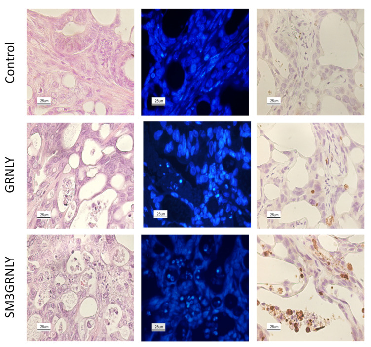
Two granulysin (GRNLY) based immunotoxins were generated, one containing the scFv of the SM3 mAb (SM3GRNLY) and the other the scFv of the AR20.5 mAb (AR20.5GRNLY). These mAb recognize different amino acid sequences of aberrantly O-glycosylated MUC1, also known as the Tn antigen, expressed in a variety of tumor cell types. We first demonstrated the affinity of these immunotoxins for their antigen using surface plasmon resonance for the purified antigen and flow cytometry for the antigen expressed on the surface of living tumor cells. The induction of cell death of tumor cell lines of different origin positive for Tn antigen expression was stronger in the cases of the immunotoxins than that induced by GRNLY alone. The mechanism of cell death induced by the immunotoxins was studied, showing that the apoptotic component demonstrated previously for GRNLY was also present, but that cell death induced by the immunotoxins included also necroptotic and necrotic components. Finally, we demonstrated the in vivo tumor targeting by the immunotoxins after systemic injection using a xenograft model of the human pancreatic adenocarcinoma CAPAN-2 in athymic mice. While GRNLY alone did not have a therapeutic effect, SM3GRNLY and AR20.5GRNLY reduced tumor volume by 42 and 60%, respectively, compared with untreated tumor-bearing mice, although the results were not statistically significant in the case of AR20.5GRNLY. Histological studies of tumors obtained from treated mice demonstrated reduced cellularity, nuclear morphology compatible with apoptosis induction and active caspase-3 detection by immunohistochemistry. Overall, our results exemplify that these immunotoxins are potential drugs to treat Tn-expressing cancers.

Keywords: MUC1; Tn antigen; granulysin; immunotoxins.


Conflict of interest statement

The use of granulysin immunotoxins as an anti-tumoral treatment is protected by the patent application PCT/ES2018/P201830768 presented to the Spanish Bureau of Patents and Brands (OEPM) on 26 July 2018; international application number PCT/EP2019/069979; number of publication WO 2020020978 A1.

Figures

Figure 1
Figure 1
Structural features of immunotoxins. Plasmid constructution and Alpha-Fold structural model of the chimeric protein of the (A) immunotoxin AR20.5GRNLY (Granulysin-AR20.5_ScFv-HisTag) and (B) SM3GRNLY (Granulysin-SM3_ScFv-HisTag). Sequence of Granulysin portion is show in green, L23 and L17 are show in grey, and fusion protein corresponding to AR20.5 and SM3 are in red and blue, respectively. AlphaFold predictions were made using the AlphaFold.ipynb v1.0 colab notebook as part of the ColabFold framework [24] and visualization of immunotoxins models was performed by PyMOL [25].
Figure 2
Figure 2
SM3GRNLY-MUC1-Tn SPR Affinity Assays. (A,B) 5700 RUs of SM3GRNLY were immobilized on the biosensor. The response was measured at different concentrations of the antigen, ranging from 0.05 µM to 500 µM. (C,D) 390 RUs of MUC1-Tn were immobilized and different concentrations of SM3GRNLY were studied. (E) KD calculation using kinetic adjustment. Colored lines correspond to the determinations with the different concentrations used in the titrations.
Figure 3
Figure 3
AR20.5GR-MUC1-Tn SPR Affinity Assays. (A,B) 6800 RUs of AR20.5GRNLY were immobilized on the biosensor, the response was measured with different concentrations of the antigen, varying from 0.07 µM to 1µM. (C) Kinetic curve of AR20.5GRNLY when the target was immobilized. Colored lines correspond to the determinations with the different concentrations used in the titrations.
Figure 4
Figure 4
Recognition of the MUC1-Tn antigen on the cell membrane of tumor cell lines. (A) expression of the MUC1-Tn antigen was analyzed on different tumor cell lines by flow cytometry using a commercial SM3 mAb conjugated with FITC. Grey histograms correspond to the isotype antibody labeling. (B) binding of the SM3GRNLY immunotoxin to tumor cell lines. Shaded histograms correspond to cells incubated with anti-His-tag antibody and FITC-conjugated goat anti-mouse IgG antibody in the absence of the immunotoxin and open histograms correspond to cells sequentially incubated at 4 °C with SM3GRNLY and the mentioned antibodies. (C) the same labelings and controls as in B were performed, comparing the binding of the SM3GRNLY (green histograms) and the AR20.5GRNLY immunotoxin (blue histograms). Images are representative of at least 3 different labeling for each cell line.
Figure 5
Figure 5
Dose-response cytotoxicity assays of GRNLY, SM3GRNLY and iSM3GRNLY on Capna-2, Jurkat and H929 cells. The cells were incubated with increasing concentrations of the recombinant proteins GRNLY (gray bars), iSM3GRNLY (red bars) or SM3GRNLY (green bars) for 24 h. GRNLY, SM3GRNLY and iSM3GRNLY induced cell death was determined by detection of PS translocation by Annexin-V-FITC labeling combined with size analysis. Results are the mean ± SD of at least three different experiments performed in triplicate. * p < 0.05, ** p < 0.01, *** p < 0.001.
Figure 6
Figure 6
Dose-response cytotoxicity assays of GRNLY and AR20.5GRNLY in CAPAN-2, MCF-7, A549, H929 and MDA-MB-231 cells. Cells were incubated with increasing concentrations of recombinant GRNLY (gray bars), iAR20.5GRNLY (dark gray bars), or AR20.5GRNLY (light gray bars) for 24 h. Cell death was determined by detecting PS exposure by Annexin-V-FITC labeling combined with size analysis (FSC-H). Results are the mean ± SD of at least three different experiments performed in triplicate for each cell line. *** p < 0.001.
Figure 7
Figure 7
Dose-response cytotoxicity assays of GRNLY and SM3GRNLY on MIA PACA-2 and MIA PACA-2 KO cells (A) or on Jurkat, Jurkat-plvTHM and Jurkat-shBak cells (B). (A) cells were incubated with increasing concentrations of recombinant GRNLY (upper panel) or of SM3GRNLY (lower panel) for 24 h; (B) cells were incubated with a 10 µM concentration of GRNLY (upper panel) or of SM3GRNLY (black bars) during the times indicated. After the incubations, cell death was determined by detecting PS exposure by Annexin-V-FITC (A) or Annexin V-DY63 (B) labeling and analysis of cell size by FSC. Results are the mean ± SD of three independent experiments; ** p < 0.01.
Figure 8
Figure 8
Study of the induction of death in JURKAT cells. Cells were preincubated for 1h in the presence or absence of 100 µM Z-VAD-fmk, 30 μM NEC-1 or 1 µM NSA alone or in combination. Subsequently, they were incubated with 10 µM of SM3GRNLY for 24 h and analyzed by flow cytometry by labeling with Annexin-V-FITC combined with the analysis of cell size in FSC-H. Representative images from two independent experiments are shown.
Figure 9
Figure 9
Systemic treatment of nude mice xenografted with CAPAN-2 cells. The mice in each group received intraperitoneal injections of GRNLY or of SM3GRNLY, as indicated, every 48 h for 10 occasions and two days after the last dose, the animals were sacrificed. Mice in the control group received PBS injections with the same schedule. Line graphics show the mean ± SD of tumor volume as a function of time in the control group and in GRNLY-treated mice (upper graphic) or in the control group and in SM3GRNLY-treated mice (lower graphic), Bar graphs correspond to the mean ± SD of the weights (upper graphic) or volumes (lower graphic) of surgically excised tumors from sacrificed mice in each experimental group, as indicated. * p < 0.05; ** p < 0.01; *** p < 0.001.
Figure 10
Figure 10
Hematoxylin-Eosin staining (left), DAPI nuclear staining (middle) and activated caspase-3 immunohistochemistry (right) of tumors derived from CAPAN-2 xenografts. Representative images of histological sections of tumors from mice treated with PBS (first row), GRNLY (second row) or SM3GRNLY (third row). Image magnification was used at 400×.

References

    1. Nath S., Mukherjee P. MUC1: A multifaceted oncoprotein with a key role in cancer progression. Trends Mol. Med. 2014;20:332–342. doi: 10.1016/j.molmed.2014.02.007. - DOI - PMC - PubMed
    1. Ju T., Otto V., Cummings R. The Tn antigen-structural simplicity and biological complexity. Angew. Chem. Int. Ed. Engl. 2011;50:1770–1791. doi: 10.1002/anie.201002313. - DOI - PMC - PubMed
    1. Martínez-Sáez N., Castro-López J., Valero-González J., Madariaga D., Compañón I., Somovilla V., Salvadó M., Asensio J., Jiménez-Barbero J., Avenoza A., et al. Deciphering the Non-Equivalence of Serine and Threonine O-Glycosylation Points: Implications for Molecular Recognition of the Tn Antigen by an anti-MUC1 Antibody. Angew. Chem. Int. Ed. Engl. 2015;54:9830–9834. doi: 10.1002/anie.201502813. - DOI - PMC - PubMed
    1. Cheever M., Allison J., Ferris A., Finn O., Hastings B., Hecht T., Mellman I., Prindiville S., Viner J., Weiner L., et al. The prioritization of cancer antigens: A national cancer institute pilot project for the acceleration of translational research. Clin. Cancer Res. 2009;15:5323–5337. doi: 10.1158/1078-0432.CCR-09-0737. - DOI - PMC - PubMed
    1. Loureiro L., Carrascal M., Barbas A., Ramalho J., Novo C., Delannoy P., Videira P. Challenges in Antibody Development against Tn and Sialyl- Tn Antigens. Biomolecules. 2015;5:1783–1809. - PMC - PubMed