. 2022 May 31;10(6):1296.
doi: 10.3390/biomedicines10061296.

Neurodynamic Treatment Promotes Mechanical Pain Modulation in Sensory Neurons and Nerve Regeneration in Rats

Affiliations

Neurodynamic Treatment Promotes Mechanical Pain Modulation in Sensory Neurons and Nerve Regeneration in Rats

Giacomo Carta et al. Biomedicines. .

Abstract

Background: Somatic nerve injuries are a rising problem leading to disability associated with neuropathic pain commonly reported as mechanical allodynia (MA) and hyperalgesia. These symptoms are strongly dependent on specific processes in the dorsal root ganglia (DRG). Neurodynamic treatment (NDT), consisting of selective uniaxial nerve repeated tension protocols, effectively reduces pain and disability in neuropathic pain patients even though the biological mechanisms remain poorly characterized. We aimed to define, both in vivo and ex vivo, how NDT could promote nerve regeneration and modulate some processes in the DRG linked to MA and hyperalgesia.

Methods: We examined in Wistar rats, after unilateral median and ulnar nerve crush, the therapeutic effects of NDT and the possible protective effects of NDT administered for 10 days before the injury. We adopted an ex vivo model of DRG organotypic explant subjected to NDT to explore the selective effects on DRG cells.

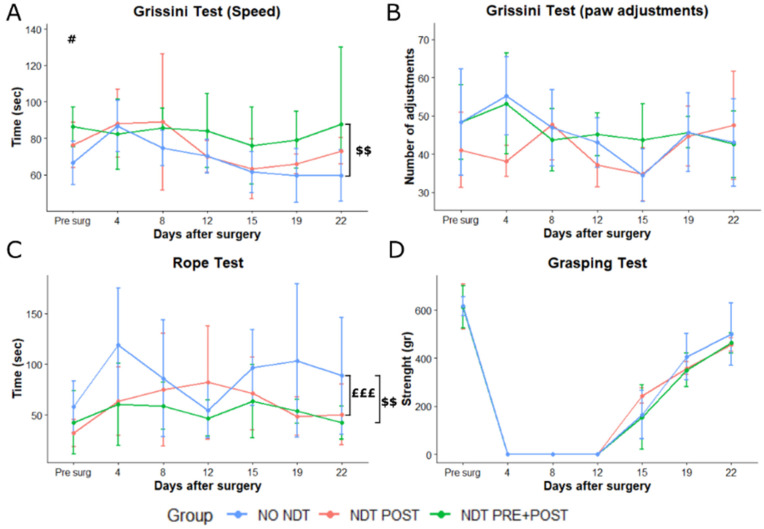
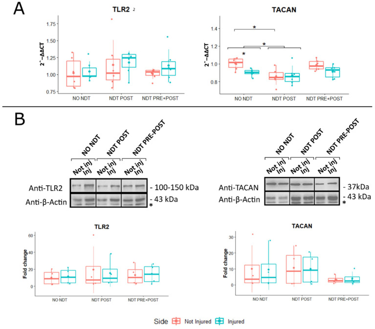
Results: Behavioural tests, morphological and morphometrical analyses, and gene and protein expression analyses were performed, and these tests revealed that NDT promotes nerve regeneration processes, speeds up sensory motor recovery, and modulates mechanical pain by affecting, in the DRG, the expression of TACAN, a mechanosensitive receptor shared between humans and rats responsible for MA and hyperalgesia. The ex vivo experiments have shown that NDT increases neurite regrowth and confirmed the modulation of TACAN.

Conclusions: The results obtained in this study on the biological and molecular mechanisms induced by NDT will allow the exploration, in future clinical trials, of its efficacy in different conditions of neuropathic pain.

Keywords: allodynia; dorsal root ganglia; gene expression; motor recovery; neurodynamic; neuropathic pain; nociceptor; non-pharmacological treatment; regeneration.


Conflict of interest statement

The authors declare no conflict of interest.

Figures

Figure 1
Figure 1
Treatment protocols and timeline of the assessments. (A) The coloured lines of the figure represent the duration of NDT protocols administrated 5 days/week, 30 sessions each day of treatment in orange (NDT POST) and green (NDT PRE + POST). (B) Assessments performed at each time point are reported in the coloured frames.
Figure 2
Figure 2
Crush injury protocol (AC). (A) After a 2 cm skin incision along the medial aspect of the long axis of the arm above the elbow, the median and ulnar nerves were isolated and exposed. (B) Median and ulnar nerves were crushed with a non-serrated clamp for 30 s. (C) The crushed area was checked to make sure there was no loss of continuity of the connective components of the nerves. Neurodynamic tensioning treatment (DH). NDT was administered to the injured side only (left side) to awake animals. The neurodynamic test from the neutral position (D) consisted of contralateral neck side flection (E), shoulder abduction and elbow extension (F), wrist extension (G), and fingers extension (H) performed until the clinician perceived resistance to mobilization.
Figure 3
Figure 3
Sensory assays. (A,B) Mechanical pain threshold and touch threshold at the paw of the median and ulnar nerve crush injured side obtained by Von Frey monofilament administration at each experimental time point (x-axis) and expressed in grams (g). (C) Pain behaviours at the Pinprick test were recorded when four consecutive pin administrations are given to the forepaw. (D) The number of withdrawals, at 22 days after injury, was recorded during 30 consecutive neurodynamic tests of the upper limb. Data are expressed as mean ± SD. The analysis compared all groups to the control group (NO NDT) with ANOVA for repeated measures (data are normally distributed with comparable variances). Differences at each time point between groups are reported * p < 0.05, *** p < 0.0001, and **** p < 0.0000, comparing the group NDT POST to the NO NDT group, or #, ### and ####, p < 0.05, p < 0.001 and p < 0.000, comparing the NDT PRE + POST to the NO NDT group. Overall differences between groups are reported £ p < 0.05 ££££ p < 0.000 comparing the NDT POST to the NO NDT group and $$$ p < 0.001 comparing the NDT PRE + POST to the NO NDT group. n = 18; 6 for each experimental group.
Figure 4
Figure 4
Motor assays. (A,B) Speed and number of paw adjustments recorded while rats were eating 7 cm grissini pieces, at different time points after nerve injury and repair. (C) Speed in climbing a 160 cm vertical rope expressed in seconds (sec). (D) Grasping strength was recorded by the device as maximal resistance to pull the grid of the device with the injured side paw. Data are expressed as mean ± SD. The analysis compared all groups to the control group (NO NDT) with ANOVA for repeated measures (data are normally distributed with comparable variances). Differences at each time point between groups are reported # p < 0.05, comparing the NDT PRE + POST to the NO NDT group. Overall differences between groups are reported £££ p < 0.001 comparing the NDT POST to the NO NDT group and $$ p < 0.01 comparing the NDT PRE + POST to the NO NDT group. Detailed data of the statistical analysis are reported in Table S2. n = 18; 6 for each experimental group.
Figure 5
Figure 5
Morphological and morphometric analysis. (A) High magnification light microscopy representative images of toluidine blue-stained semi-thin cross-sections of uninjured median nerves and injured median nerves after 24 days from the lesion, for each treatment. Scale bar: 20 µm. (BH) Stereological assessment of morphological changes during nerve regeneration. (B) Number of myelinated fibres; (C) myelinated fibre density; (D) axon diameters; (E); fibre diameters; (F) myelin thickness; (G) axon diameter/fibre diameter ratio (g-ratio); (H) intraneural collagen area. Data are expressed as mean ± SD. The analysis compared all groups and sides to the control group (NO NDT) with a Two-way ANOVA (data are normally distributed with comparable variances). Differences are reported * p < 0.05, ** p < 0.01, and **** p < 0.0000. n = 18; 6 for each experimental group.
Figure 6
Figure 6
Gene and protein expression analysis of markers linked to mechanical allodynia and apoptosis expressed in the DRG. Panel (A): Beneficial or side effects induced by the neurodynamic protocols on DRG were assessed adopting the relative quantification (2-ΔΔCt) of genes by qRT-PCR. Data normalization was performed considering TATA-box binding protein (TBP) as a housekeeping gene. All data were calibrated to NO NDT Not Injured samples. Panel (B): A representative Western blot is shown; actin was used as a loading control. Asterisks (*) in panel B identify unspecific bands. Values in the graphics are expressed as mean ± SD. Respectively, two-way and one-way ANOVA were carried out (data are normally distributed with comparable variances). * p < 0.05. n = 18; 6 for each experimental group.
Figure 7
Figure 7
Effects of neurodynamic treatment on cell morphology, gene and protein expression on dorsal root ganglia (DRG) explants. Panel (A): Representative images of rat organotypic dorsal root ganglia neurons stained with βIII-tubulin reported for each type of experimental protocol. Scale bar: 400 µm. Panel (B): Quantitative analysis of the distance of the longest neurite (Dmax), the maximum number of neurites (Nmax), and the Sholl critical value, defined as the distance from the organotypic culture centre. Values in the graphics are expressed as mean ± SD. Panel (C): Gene expression analysis of markers linked to mechanical allodynia and neuropathic pain. Beneficial or side effects induced by the neurodynamic protocols on dorsal root ganglia explants were assessed adopting the relative quantification analysis (2−ΔΔCt) of genes by qRT-PCR. Data normalization was performed considering TATA-box binding protein (TBP) as a housekeeping gene. All data were calibrated to CTR OUT sample). Panel (D): BAX and Bcl-xL protein expression in DRG explants. Protocols are described as follows: not treated (CTR IN), sham-treated (CTR OUT), and treated (NDT) with 30 repetitions of neurodynamic treatment. Experiments were carried out in a biological octuplicate (n = 21 for a technical replicate). Asterisks (*) in panel (D) identify unspecific bands. Values in the graphics are expressed as mean ± SD. For normally distributed data with comparable variances One-way ANOVA was carried out, while Kruskal–Wallis test was used for nonparametric data; asterisks show statistically significant differences with CTR OUT (sham sample); * p < 0.05, and ** p < 0.01.

Similar articles

Cited by

References

    1. Hecke O.V., Austin S.K., Khan R.A., Smith B.H., Torrance N. Neuropathic pain in the general population: A systematic review of epidemiological studies. Pain. 2014;155:654–662. doi: 10.1016/j.pain.2013.11.013. - DOI - PubMed
    1. Smith B.H., Harry L.H., Hébert H.L., Veluchamy A. Neuropathic pain in the community: Prevalence, impact, and risk factors. Pain. 2020;161:S127–S137. doi: 10.1097/j.pain.0000000000001824. - DOI - PubMed
    1. Machado-Duque M.E., Gaviria-Mendoza A., Machado-Alba J.E., Castaño N. Evaluation of Treatment Patterns and Direct Costs Associated with the Management of Neuropathic Pain. Pain Res. Manag. 2020;2020:8. doi: 10.1155/2020/9353940. - DOI - PMC - PubMed
    1. Loeser J.D., Cahana A. Pain medicine versus pain management: Ethical dilemmas created by contemporary medicine and business. Clin. J. Pain. 2013;29:311–316. doi: 10.1097/AJP.0b013e3182516e64. - DOI - PubMed
    1. Darnall B. To treat pain, study people in all their complexity. Nature. 2018;557:7–8. doi: 10.1038/d41586-018-04994-5. - DOI - PubMed

LinkOut - more resources