. 2022 Jun 1;10(6):1297.
doi: 10.3390/biomedicines10061297.

Galanin and Neuropeptide Y Interaction Enhances Proliferation of Granule Precursor Cells and Expression of Neuroprotective Factors in the Rat Hippocampus with Consequent Augmented Spatial Memory

Affiliations

Galanin and Neuropeptide Y Interaction Enhances Proliferation of Granule Precursor Cells and Expression of Neuroprotective Factors in the Rat Hippocampus with Consequent Augmented Spatial Memory

Marina Mirchandani-Duque et al. Biomedicines. .

Abstract

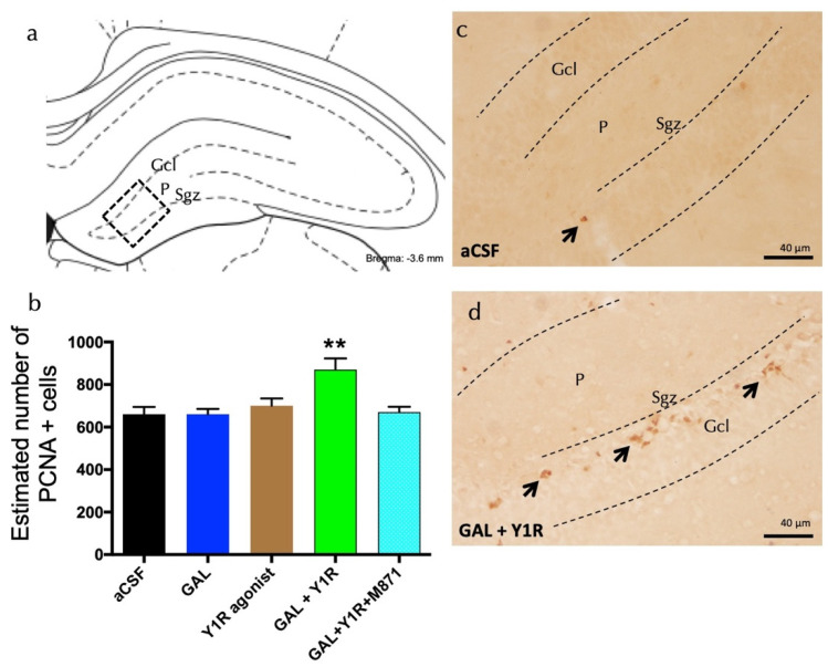
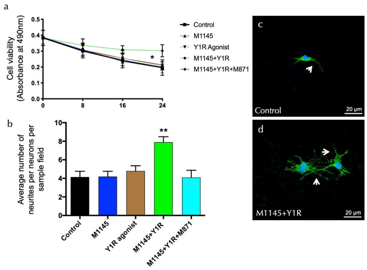
Dysregulation of hippocampal neurogenesis is linked to several neurodegenereative diseases, where boosting hippocampal neurogenesis in these patients emerges as a potential therapeutic approach. Accumulating evidence for a neuropeptide Y (NPY) and galanin (GAL) interaction was shown in various limbic system regions at molecular-, cellular-, and behavioral-specific levels. The purpose of the current work was to evaluate the role of the NPY and GAL interaction in the neurogenic actions on the dorsal hippocampus. We studied the Y1R agonist and GAL effects on: hippocampal cell proliferation through the proliferating cell nuclear antigen (PCNA), the expression of neuroprotective and anti-apoptotic factors, and the survival of neurons and neurite outgrowth on hippocampal neuronal cells. The functional outcome was evaluated in the object-in-place task. We demonstrated that the Y1R agonist and GAL promote cell proliferation and the induction of neuroprotective factors. These effects were mediated by the interaction of NPYY1 (Y1R) and GAL2 (GALR2) receptors, which mediate the increased survival and neurites' outgrowth observed on neuronal hippocampal cells. These cellular effects are linked to the improved spatial-memory effects after the Y1R agonist and GAL co-injection at 24 h in the object-in-place task. Our results suggest the development of heterobivalent agonist pharmacophores, targeting Y1R-GALR2 heterocomplexes, therefore acting on the neuronal precursor cells of the DG in the dorsal hippocampus for the novel therapy of neurodegenerative cognitive-affecting diseases.

Keywords: BDNF; Bcl-2; dorsal hippocampus; galanin 2 receptor; neurodegenerative diseases; neurogenesis; neuropeptide Y1 receptor; neuroprotection; receptor–receptor interaction; spatial memory.


Conflict of interest statement

The authors declare no conflict of interest.

Figures

Figure 1
Figure 1
Co-administration of galanin and Y1R agonists increases cell proliferation in the dentate gyrus of adult rats. Proliferating cell nuclear antigen immunolabeling (PCNA+) in the dentate gyrus of the dorsal hippocampus, after the intracerebroventricular (icv) administration of galanin (GAL) and Y1R receptor agonists, either alone or in combination with or without the GAL 2 receptor antagonist (M871). (a,d) The majority of the PCNA-IR cells were located in the sub-granular zone (Sgz) of the dentate gyrus at the border between the granular cell layer (Gcl) and the polymorphic layer (P) of the dentate gyrus in the dorsal hippocampus. They appeared as groups of 3–4 cells (Bregma: −3.6 mm, according to the Paxinos and Watson stereotaxic atlas [33]). (b) Quantification of total PCNA-IR cells in the dorsal hippocampal dentate gyrus. Data represent mean ± SEM, showing the differences between groups after the injections of aCSF, GAL, the Y1R agonist [Leu31,Pro34]NPY, or the co-administration of both substances with or without M871. GAL and the Y1R agonist co-administration augmented the number of PCNA+ cells in the dorsal hippocampus compared to the lack of effects of the two peptides alone and the aCSF group. Additionally, this effect was blocked by the GALR2 antagonist M871. ** p < 0.01 vs. the rest of the groups according to one-way ANOVA followed by Newman–Keuls post-hoc test. N = 4 in each group. Statistical values are presented in Supplementary Table S1. GAL and Y1R agonist co-injection (d) increased the PCNA-IR cells in Sgz in the dentate gyrus compared with the control group (c). Arrows indicate examples of clusters of PCNA+ nerve cells. Dashed lines outline the Gcl of the dentate gyrus. Abbreviations: aCSF = Control (artificial cerebrospinal fluid), GAL = galanin (3 nmol), Y1R agonist = Y1R receptor agonist [Leu31, Pro34]NPY (3 nmol), GAL + Y1R = co-administration of GAL and Y1R, GAL + Y1R + M871 = co-administration of GAL, Y1R, and GALR2 antagonist M871 (3 nmol).
Figure 2
Figure 2
Galanin and the Y1R agonist effects on hippocampal brain-derived neurotrophic factor-immunoreactive (BDNF-IR) cells of the dentate gyrus (DG) hippocampal region. (a) BDNF-IR cells were located mainly in the sub-granular zone (Sgz) of the dentate gyrus at the border of the granular cell layer (Gcl), and some scattered cells were found in the polymorphic layer (P) of the dentate gyrus in the dorsal hippocampus (Bregma: −3.6 mm, according to the Paxinos and Watson [33] stereotaxic atlas). (b) Quantitative morphometric analysis of BDNF-IR cells of the DG. GAL and Y1R agonist icv co-administration significantly increased BDNF-IR cells in the dorsal DG. This effect was counteracted in the presence of the GALR2 antagonist M871. * p < 0.05 vs. Y1R agonist and GAL + Y1R + M871; ** p < 0.01 vs. aCSF and GAL according to one-way ANOVA followed by Newman–Keuls post-hoc test. The vertical lines from the horizontal line above the bars indicate the inter-group comparisons. Data are expressed as mean ± SEM, n = 4. Statistical values are presented in Supplementary Table S1. (c,d) Representative microphotographs of the significant increment in the BDNF-positive cells in the DG after GAL and Y1R agonist co-injection (d) compared with the control group (c). The cells in red are BDNF-positive using confocal laser microscopy. White arrows point to BDNF-IR cells. Dashed lines outline the Gcl of the dentate gyrus. The nuclei are shown in blue by DAPI. Abbreviations: aCSF = Control (artificial cerebrospinal fluid), GAL = galanin (3 nmol), Y1R agonist = Y1R receptor agonist [Leu31, Pro34]NPY (3 nmol), GAL + Y1R = co-administration of GAL and Y1R, GAL + Y1R + M871 = co-administration of GAL, Y1R, and GALR2 antagonist M871 (3 nmol).
Figure 3
Figure 3
Dentate gyrus (DG) hippocampal anti-apoptotic Bcl-2-immunoreactive cells (Bcl-2-IR) after galanin and the Y1R agonist co-administration. (a) Bcl-2-IR cells were located in the sub-granular zone (Sgz) of the dentate gyrus at the border between the granular cell layer (Gcl) and the polymorphic layer (P) of the dentate gyrus in the dorsal hippocampus (Bregma: −3.6 mm, according to the Paxinos and Watson [33] stereotaxic atlas). (b) Quantitative morphometric analysis of Bcl-2-IR cells of the DG. GAL and Y1R agonist icv co-administration significantly increased Bcl-2-IR cells in the dorsal DG. This effect was blocked by treatment with the GALR2 antagonist M871. * p < 0.05 vs. the rest of the groups according to one-way ANOVA followed by Newman–Keuls post-hoc test. Data are expressed as mean ± SEM, n = 4. Statistical values are presented in Supplementary Table S1. (c,d) Representative microphotographs of the significant increase in the Bcl-2-positive cells in the DG after GAL and Y1R agonist co-injection (d) compared with the control group (c). The cells in green are BDNF-positive using confocal laser microscopy. White arrows point to Bcl-2-IR cells. Dashed lines outline the Gcl of the dentate gyrus. The nuclei are shown in blue by DAPI. Abbreviations: aCSF = Control (artificial cerebrospinal fluid), GAL = galanin (3 nmol), Y1R agonist = Y1R receptor agonist [Leu31-Pro34]NPY (3 nmol), GAL + Y1R = co-administration of GAL and Y1R, GAL + Y1R + M871 = co-administration of GAL, Y1R, and GALR2 antagonist M871 (3 nmol).
Figure 4
Figure 4
Evaluation of survival and neurite outgrowth on hippocampal neuronal cells. (a) Cell viability was determined by the MTS assay (CellTiter 96A Aqueous One Solution Cell Proliferation Assay (Promega)) with hippocampal neurons after treatment with the galanin receptor 2 agonist (M1145, 100 nM) and Y1R receptor agonist (100 nM), either alone or in combination with or without the GAL 2 receptor antagonist (M871, 1 µM). Cells (20,000 per well) were seeded, and after seven days, the cells were incubated for 0, 8, 16, and 24 h in triplicates with the different groups. Blanks (medium only plus CellTiter 96A Aqueous One Solution Reagent) were subtracted from the value measured for each well incubated with the groups. M1145 and Y1R agonists’ incubation significantly increased neuronal survival in the dorsal DG. This effect was blocked by treatment with the GALR2 antagonist M871. * p < 0.05, 24 h after M1145 and Y1R agonist co-stimulation compared with every group according to Student’s unpaired t-test statistical analysis. Statistical values are presented in Supplementary Table S1. (bd) GALR2 and Y1R agonists’ modulation of neurites’ outgrowth. Primary hippocampal neurons were treated for 24 h without (control) or with M1145 (100 nM) and/or Y1R agonists (100 nM) with or without the GAL 2 receptor antagonist (M871, 1 µM). The numbers of neurites per cell were determined after immunofluorescent labeling of neurons and neuronal nuclei (Pan Neuronal Marker (ABN2300)/neuronal nuclei (DAPI)). Quantification is shown in Figure 4b, where the data are presented as mean ± SEM. The combined group is significantly different from the rest of the groups (** p < 0.01 vs. the rest of the groups according to one-way ANOVA followed by Newman–Keuls post-hoc test). Statistical values are presented in Supplementary Table S1. Representative microphotographs of the significant increase in the number of neurites in the hippocampal cells after M1145 and Y1R agonist treatment (d) compared with the control group (c). The cells in green are hippocampal neuron-positive using confocal laser microscopy. White arrows point to neurite extensions. The nuclei are counterstained in blue by DAPI. Abbreviations: Control = Culture medium, M1145 = galanin 2 receptor agonist (100 nM), Y1R agonist = Y1R receptor agonist [Leu31-Pro34]NPY (100 nM), M1145 + Y1R = co-administration of M1145 and Y1R, M1145 + Y1R + M871 = co-administration of M1145, Y1R, and GALR2 antagonist (1 µM).
Figure 5
Figure 5
Spatial memory assessment after GAL and the Y1R agonist alone and combined in the object-in-place memory task. (a) Schematic representation of the trials completed in the object-in-place task. The animals performed the task in three phases, divided 24 h from each other, where they explored freely for ten minutes in the habituation phase without objects, three minutes in the training phase with four different objects, and finally, three minutes in the test phase with two of the objects with the exchanged position. To achieve memory consolidation, the pharmacological treatments were administered intracerebroventricularly (icv) to the different groups of animals 24 h before the testing phase. (b) Performance on the object-in-place task showing the ability of rats to discriminate the exchanged objects at 24 h post-training after the icv administration of GAL in combination with the Y1R agonist. An improvement in the object-in-place performance was observed after GAL and the Y1 agonist co-administration following a 24 h delay. Besides, this effect is counteracted by the GAL 2 receptor (GALR2) antagonist M871. Data are presented as the mean ± SEM of the discrimination ratio on the test phase. n = 6 animals in each group. * p < 0.05 vs. Y1R agonist and GAL + Y1R + M871; ** p < 0.01 vs. aCSF; *** p < 0.001 vs. GAL according to one-way ANOVA (F4, 25 = 3.56) followed by Newman–Keuls post-hoc test. Statistical values are presented in Supplementary Table S1. Abbreviations: aCSF = Control (artificial cerebrospinal fluid), GAL = galanin (3 nmol), Y1R agonist = Y1R receptor agonist [Leu31-Pro34]NPY (3 nmol), GAL + Y1R = co-administration of GAL and Y1R, GAL + Y1R + M871 = co-administration of GAL, Y1R, and GALR2 antagonist M871 (3 nmol).

Similar articles

Cited by

References

    1. Baptista P., Andrade J.P. Adult Hippocampal Neurogenesis: Regulation and Possible Functional and Clinical Correlates. Front. Neuroanat. 2018;12:44. doi: 10.3389/fnana.2018.00044. - DOI - PMC - PubMed
    1. Spalding K.L., Bergmann O., Alkass K., Bernard S., Salehpour M., Huttner H.B., Bostrom E., Westerlund I., Vial C., Buchholz B.A., et al. Dynamics of hippocampal neurogenesis in adult humans. Cell. 2013;153:1219–1227. doi: 10.1016/j.cell.2013.05.002. - DOI - PMC - PubMed
    1. Fanselow M.S., Dong H.W. Are the dorsal and ventral hippocampus functionally distinct structures? Neuron. 2010;65:7–19. doi: 10.1016/j.neuron.2009.11.031. - DOI - PMC - PubMed
    1. Kheirbek M.A., Drew L.J., Burghardt N.S., Costantini D.O., Tannenholz L., Ahmari S.E., Zeng H., Fenton A.A., Hen R. Differential control of learning and anxiety along the dorsoventral axis of the dentate gyrus. Neuron. 2013;77:955–968. doi: 10.1016/j.neuron.2012.12.038. - DOI - PMC - PubMed
    1. Tanti A., Belzung C. Neurogenesis along the septo-temporal axis of the hippocampus: Are depression and the action of antidepressants region-specific? Neuroscience. 2013;252:234–252. doi: 10.1016/j.neuroscience.2013.08.017. - DOI - PubMed