

Characterization of Epigenetic and Molecular Factors in Endometrium of Females with Infertility
- PMID: 35740346
- PMCID: PMC9219839
- DOI: 10.3390/biomedicines10061324
Characterization of Epigenetic and Molecular Factors in Endometrium of Females with Infertility
Abstract
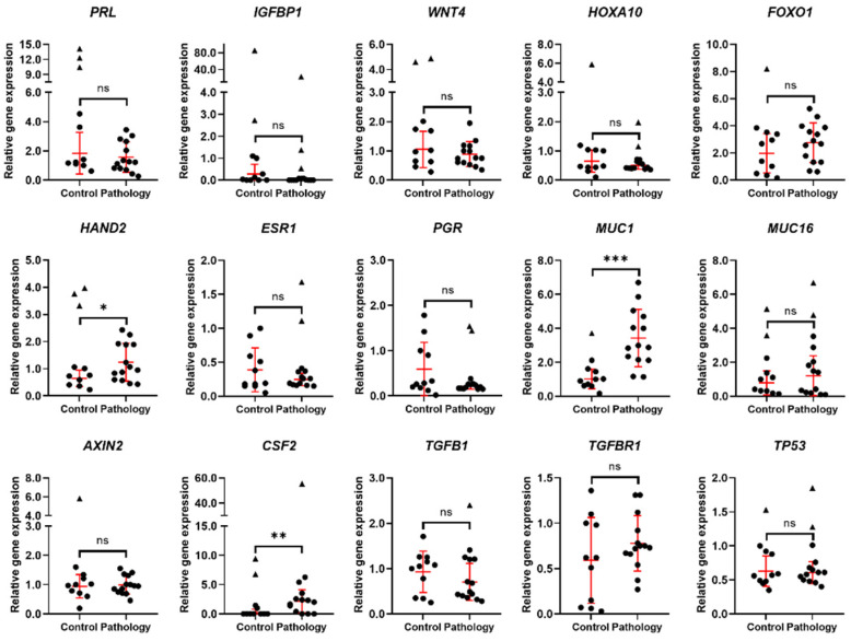
Infertility is one of the most rapidly increasing global health concerns of the 21st century. Embryo quality and endometrial thickness and receptivity are the main factors for successful embryo implantation and pregnancy development. Nevertheless, until now, there has been a lack of understanding about the regulation of human endometrium function and its structure. This raises the demand for more research of the human endometrium in these fields. In our study, we analyzed the genetic and epigenetic changes of endometrial tissue's samples isolated from females admitted for treatment due to male infertility and females diagnosed with reproductive pathologies, who are preparing for assisted reproductive technologies procedures. Using real-time polymerase chain reaction method, we demonstrated that endometrium of females with reproductive pathology has significantly upregulated decidualization related genes HAND2, MUC1, CSF2, increased expression of angiogenesis related gene PDGFA, and increases of overall immune response and inflammation-related genes expression with significant changes of RELA and CXCL10 genes expression. Females with reproductive pathology have altered endometrium epigenetic regulation since expression of miRNAs-specifically, miRNA-34a, miRNA-223, and miRNA-125b-is lower in endometrium of females with reproductive pathology. Our findings suggest that the potential changes in genetic and epigenetic profile of endometrium from females with reproductive pathology could enrich the knowledge in the field of core biological knowledge and treatment of reproductive impairments.
Keywords: endometrial tissue; endometrium; infertility; reproduction; reproductive disorders; reproductive pathology.
Conflict of interest statement
The authors declare no conflict of interest.
Figures


Similar articles
-
[Differential expression of microRNA in eutopic endometrium tissue during implantation window for patients with endometriosis related infertility].Zhonghua Fu Chan Ke Za Zhi. 2016 Jun 25;51(6):436-41. doi: 10.3760/cma.j.issn.0529-567X.2016.06.007. Zhonghua Fu Chan Ke Za Zhi. 2016. PMID: 27356479 Chinese.
-
Gradient Boosting Machine Learning Model for Defective Endometrial Receptivity Prediction by Macrophage-Endometrium Interaction Modules.Front Immunol. 2022 May 6;13:842607. doi: 10.3389/fimmu.2022.842607. eCollection 2022. Front Immunol. 2022. PMID: 35603216 Free PMC article.
-
Development of a new comprehensive and reliable endometrial receptivity map (ER Map/ER Grade) based on RT-qPCR gene expression analysis.Hum Reprod. 2018 Feb 1;33(2):220-228. doi: 10.1093/humrep/dex370. Hum Reprod. 2018. PMID: 29315421
-
Reproductive ecology and the endometrium: physiology, variation, and new directions.Am J Phys Anthropol. 2009;140 Suppl 49:137-54. doi: 10.1002/ajpa.21188. Am J Phys Anthropol. 2009. PMID: 19890864 Review.
-
Endometrial receptivity and pregnancy outcome.J Matern Fetal Neonatal Med. 2022 Jul;35(13):2591-2605. doi: 10.1080/14767058.2020.1787977. Epub 2020 Aug 2. J Matern Fetal Neonatal Med. 2022. PMID: 32744104 Review.
Cited by
-
Palmitic Acid Upregulates CD96 Expression to Mediate Maternal-Foetal Interface Immune Tolerance by Inhibiting Cytotoxic Activity and Promoting Adhesion Function in Human Decidual Natural Killer Cells.Bioengineering (Basel). 2023 Aug 25;10(9):1008. doi: 10.3390/bioengineering10091008. Bioengineering (Basel). 2023. PMID: 37760110 Free PMC article.
-
Investigation of hub ferroptosis-related genes and the immune landscape in recurrent pregnancy loss and unexplained infertility using bioinformatics analysis.Ann Transl Med. 2023 Mar 15;11(5):209. doi: 10.21037/atm-23-97. Epub 2023 Mar 2. Ann Transl Med. 2023. PMID: 37007565 Free PMC article.
-
Estrogen-Regulated Proline-Rich Acidic Protein 1 in Endometrial Epithelial Cells Affects Embryo Implantation by Regulating Mucin 1 in Mice.Biomolecules. 2025 Jun 11;15(6):852. doi: 10.3390/biom15060852. Biomolecules. 2025. PMID: 40563492 Free PMC article.
-
Hysteroscopic Endometrial Fundal Incision in Oocyte Recipients before Embryo Transfer May Improve Reproductive Outcomes: A Prospective Study.Int J Fertil Steril. 2023 Nov 7;18(1):40-44. doi: 10.22074/ijfs.2023.560746.1354. Int J Fertil Steril. 2023. PMID: 38041458 Free PMC article.
References
LinkOut - more resources
Full Text Sources
Research Materials
Miscellaneous
