. 2022 Jun 7;10(6):1345.
doi: 10.3390/biomedicines10061345.

Inhibiting Transglutaminase 2 Mediates Kidney Fibrosis via Anti-Apoptosis

Affiliations

Inhibiting Transglutaminase 2 Mediates Kidney Fibrosis via Anti-Apoptosis

Jong-Joo Moon et al. Biomedicines. .

Abstract

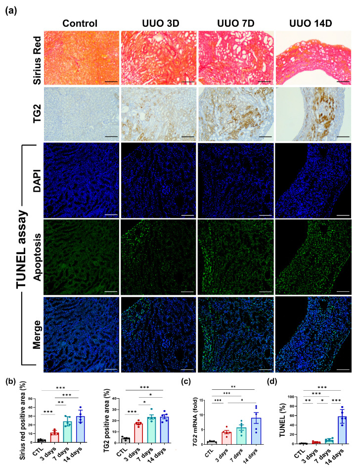
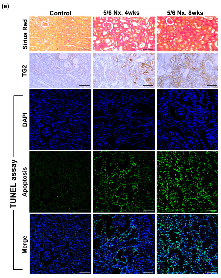
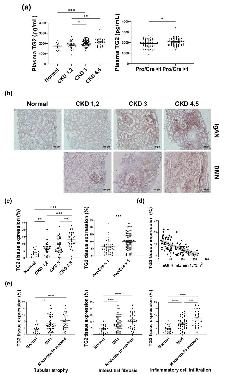
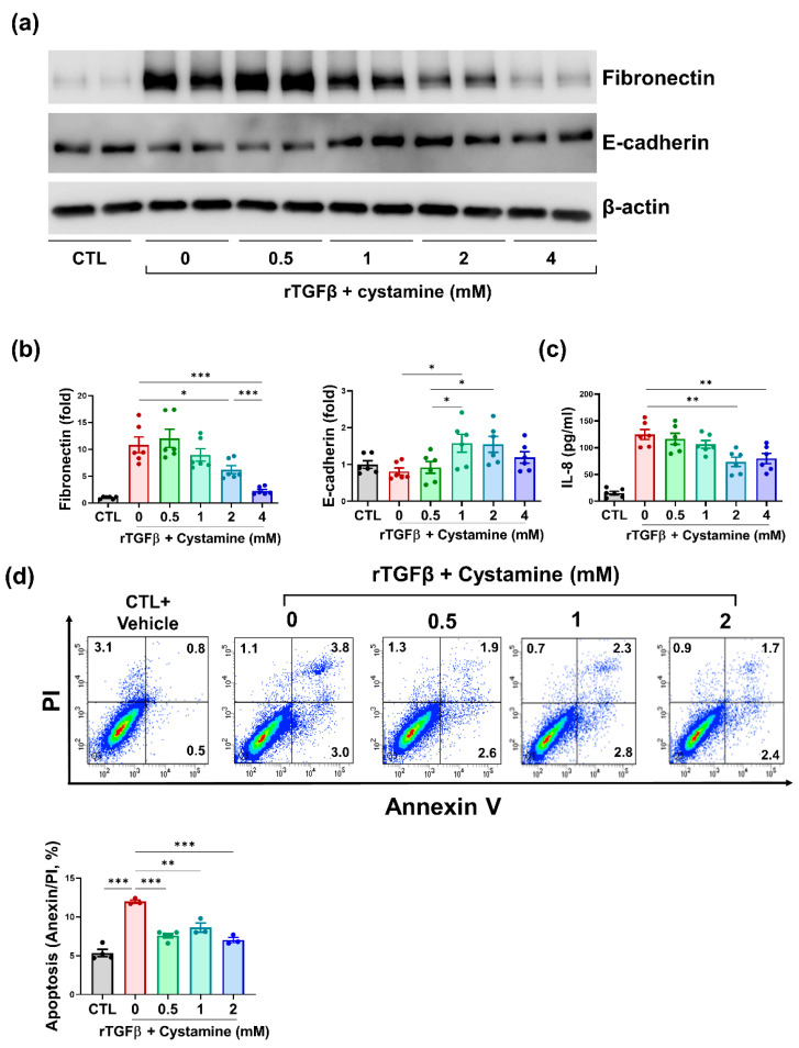
Transglutaminase 2 (TG2) is a calcium-dependent transamidating acyltransferase enzyme of the protein-glutamine γ-glutamyltransferase family implicated in kidney injury. In this study, we identified associations between TG2 and chronic kidney disease (CKD) identified by visualizing TG2 in kidney biopsy samples derived from CKD patients using immunohistochemistry and measuring the plasma TG2 concentrations. Our study revealed a connection between TG2 and the pathological markers of kidney disease. We showed high plasma TG2 levels in samples from patients with advanced CKD. In addition, we observed an increase in TG2 expression in tissues concomitant with advanced CKD in human samples. Moreover, we investigated the effect of TG2 inhibition on kidney injury using cystamine, a well-known competitive inhibitor of TG2. TG2 inhibition reduced apoptosis and accumulation of extracellular molecules (ECM) such as fibronectin and pro-inflammatory cytokine IL-8. Collectively, the increased expression of TG2 that was observed in advanced CKD, hence inhibiting TG2 activity, could protect kidney cells from ECM molecule accumulation, apoptosis, and inflammatory responses, thereby preventing kidney fibrosis.

Keywords: apoptosis; chronic kidney disease; cystamine; fibrosis; transglutaminase 2.


Conflict of interest statement

The authors have no conflict of interest to declare.

Figures

Figure 1
Figure 1
Increased TG2 levels were accompanied by pathological aggravation in UUO and 5/6 nephrected (Nx.) rodent models. UUO kidneys were evaluated thrice, on days 3, 7, and 14, after the establishment of the UUO model. (a) Representative images of UUO kidneys stained with Sirius red, immunohistochemical staining for TG2, and TUNEL assay on days 3, 7, and 14. (b) Semi-quantification results for Sirius red and TG2-stained UUO kidney. (c) Changes in TG2 mRNA abundance after 3, 7, and 14 days from UUO surgery. (d) Percentage of TUNEL positive area on days 3, 7, and 14. (e) In the 5/6 Nx. model, kidney tissue-expressed TG2 levels were evaluated twice at 4 and 8 weeks. Representative images of 5/6 Nx. kidneys stained with Sirius red, immunostained with TG2, and TUNEL assay. (f) Semi-quantification results for TG2-stained 5/6 Nx. Kidney. (g) Apoptosis was assessed twice at 4 and 8 weeks using the TUNEL assay. (Scale bar = 100μm, * p <0.05, ** p < 0.01, *** p < 0.001).
Figure 1
Figure 1
Increased TG2 levels were accompanied by pathological aggravation in UUO and 5/6 nephrected (Nx.) rodent models. UUO kidneys were evaluated thrice, on days 3, 7, and 14, after the establishment of the UUO model. (a) Representative images of UUO kidneys stained with Sirius red, immunohistochemical staining for TG2, and TUNEL assay on days 3, 7, and 14. (b) Semi-quantification results for Sirius red and TG2-stained UUO kidney. (c) Changes in TG2 mRNA abundance after 3, 7, and 14 days from UUO surgery. (d) Percentage of TUNEL positive area on days 3, 7, and 14. (e) In the 5/6 Nx. model, kidney tissue-expressed TG2 levels were evaluated twice at 4 and 8 weeks. Representative images of 5/6 Nx. kidneys stained with Sirius red, immunostained with TG2, and TUNEL assay. (f) Semi-quantification results for TG2-stained 5/6 Nx. Kidney. (g) Apoptosis was assessed twice at 4 and 8 weeks using the TUNEL assay. (Scale bar = 100μm, * p <0.05, ** p < 0.01, *** p < 0.001).
Figure 2
Figure 2
TG2 levels were increased with deterioration of kidney function and pathological aggravation in patient samples. (a) The concentration of TG2 was evaluated in the plasma of CKD patients. (b) Representative images of immunohistochemical staining for TG2. (c) Differences in the tissue expression level of TG2 according to kidney function markers, including CKD stage and urine protein/creatinine ratio. (d) Association between TG2 expression and eGFR. (e) Differences in TG2 expression levels according to tubular pathology (* p < 0.05, ** p < 0.01, *** p < 0.001).
Figure 3
Figure 3
Cystamine-mediated inhibition of TG2 protected hTECs from rTGFβ-induced injury. (a) Representative images of western blots for fibronectin and E-cadherin. (b) Semi-quantification results for fibronectin and E-cadherin. (c) Concentration of IL-8 in cell culture media. (d) Evaluation for cell death conducted using Annexin V/PI assay in hTECs 48 h after rTGFβ induction (* p < 0.05, ** p < 0.01, *** p < 0.001).

Similar articles

Cited by

References

    1. Jha V., Garcia-Garcia G., Iseki K., Li Z., Naicker S., Plattner B., Saran R., Wang A.Y.-M., Yang C.-W. Chronic kidney disease: Global dimension and perspectives. Lancet. 2013;382:260–272. doi: 10.1016/S0140-6736(13)60687-X. - DOI - PubMed
    1. Hill N.R., Fatoba S.T., Oke J.L., Hirst J.A., O’Callaghan C.A., Lasserson D.S., Hobbs F.D. Global Prevalence of Chronic Kidney Disease-A Systematic Review and Meta-Analysis. PLoS ONE. 2016;11:e0158765. doi: 10.1371/journal.pone.0158765. - DOI - PMC - PubMed
    1. Ruiz-Ortega M., Rayego-Mateos S., Lamas S., Ortiz A., Rodrigues-Diez R.R. Targeting the progression of chronic kidney disease. Nat. Rev. Nephrol. 2020;16:269–288. doi: 10.1038/s41581-019-0248-y. - DOI - PubMed
    1. Min B., Chung K.C. New insight into transglutaminase 2 and link to neurodegenerative diseases. BMB Rep. 2018;51:5–13. doi: 10.5483/BMBRep.2018.51.1.227. - DOI - PMC - PubMed
    1. Di Sabatino A., Vanoli A., Giuffrida P., Luinetti O., Solcia E., Corazza G.R. The function of tissue transglutaminase in celiac disease. Autoimmun. Rev. 2012;11:746–753. doi: 10.1016/j.autrev.2012.01.007. - DOI - PubMed

LinkOut - more resources