

Exploring Anatomo-Morphometric Characteristics of Infrapatellar, Suprapatellar Fat Pad, and Knee Ligaments in Osteoarthritis Compared to Post-Traumatic Lesions
- PMID: 35740391
- PMCID: PMC9220326
- DOI: 10.3390/biomedicines10061369
Exploring Anatomo-Morphometric Characteristics of Infrapatellar, Suprapatellar Fat Pad, and Knee Ligaments in Osteoarthritis Compared to Post-Traumatic Lesions
Abstract
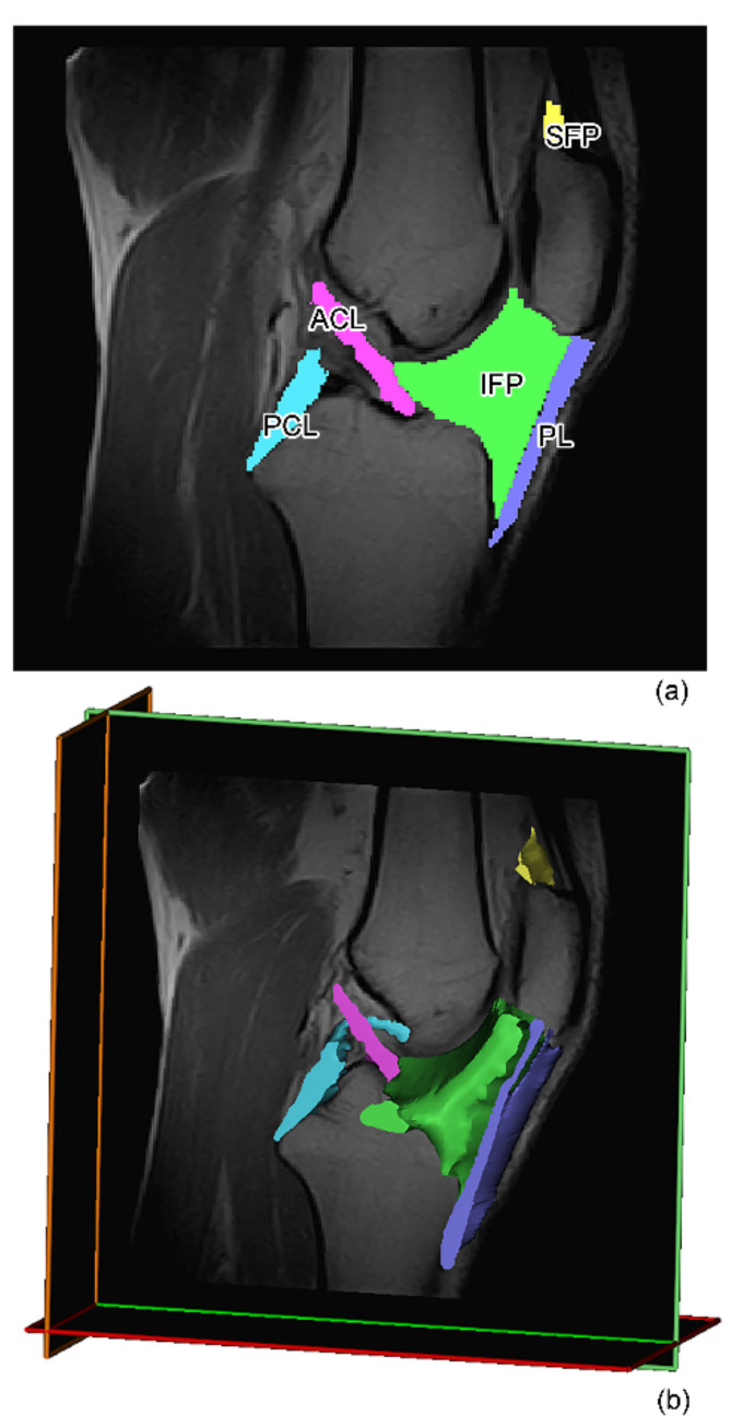
Several studies have investigated cartilage degeneration and inflammatory subchondral bone and synovial membrane changes using magnetic resonance (MR) in osteoarthritis (OA) patients. Conversely, there is a paucity of data exploring the role of knee ligaments, infrapatellar fat pad (IFP), and suprapatellar fat pad (SFP) in knee OA compared to post-traumatic cohorts of patients. Therefore, the aim of this study was to analyze the volumetric and morphometric characteristics of the following joint tissues: IFP (volume, surface, depth, femoral and tibial arch lengths), SFP (volume, surface, oblique, antero−posterior, and cranio−caudal lengths), anterior (ACL) and posterior cruciate ligament (PCL) (volume, surface, and length), and patellar ligament (PL) (volume, surface, arc, depth, and length). Eighty-nine MR images were collected in the following three groups: (a) 32 patients with meniscal tears, (b) 29 patients with ACL rupture (ACLR), and (c) 28 patients affected by end-stage OA. Volume, surface, and length of both ACL and PCL were determined in groups a and c. A statistical decrease of IFP volume, surface, depth, femoral and tibial arch lengths was found in end-stage OA compared to patients with meniscal tear (p = 0.002, p = 0.008, p < 0.0001, p = 0.028 and p < 0.001, respectively) and patients with ACLR (p < 0.0001, p < 0.0001, p = 0.008 and p = 0.011, respectively). An increment of volume and surface SFP was observed in group b compared to both groups a and c, while no differences were found in oblique, antero−posterior, and cranio−caudal lengths of SFP among the groups. No statistical differences were highlighted comparing volume, surface, arc, and length of PL between the groups, while PL depth was observed to be decreased in end-OA patients compared with meniscal tear patients (p = 0.023). No statistical differences were observed comparing ACL and PCL lengths between patients undergoing meniscectomy and TKR. Our study confirms that IFP MR morphometric characteristics are different between controls and OA, supporting an important role of IFP in OA pathology and progression in accordance with previously published studies. In addition, PL depth changes seem to be associated with OA pathology. Multivariate analysis confirmed that OA patients had a smaller IFP compared to patients with meniscal tears, confirming its involvement in OA.
Keywords: anterior cruciate ligament rupture; infrapatellar fat pad; knee; magnetic resonance; meniscal tear; osteoarthritis; patellar ligament; posterior cruciate ligament; segmentation; suprapatellar fat pad.
Conflict of interest statement
The authors declare no conflict of interest. The funders had no role in the design of the study; in the collection, analyses, or interpretation of data; in the writing of the manuscript, or in the decision to publish the results.
Figures


References
-
- Callahan L.F., Ambrose K.R., Albright A.L., Altpeter M., Golightly Y.M., Huffman K.F., Nelson A.E., Weisner S.E. Public Health Interventions for Osteoarthritis—Updates on the Osteoarthritis Action Alliance’s efforts to address the 2010 OA Public Health Agenda Recommendations. Clin. Exp. Rheumatol. 2019;37((Suppl. S120)):31–39. - PubMed
-
- Belluzzi E., Stocco E., Pozzuoli A., Granzotto M., Porzionato A., Vettor R., De Caro R., Ruggieri P., Ramonda R., Rossato M., et al. Contribution of Infrapatellar Fat Pad and Synovial Membrane to Knee Osteoarthritis Pain. BioMed Res. Int. 2019;2019:6390182. doi: 10.1155/2019/6390182. - DOI - PMC - PubMed
LinkOut - more resources
Full Text Sources
