. 2022 Jun 10;10(6):1382.
doi: 10.3390/biomedicines10061382.

Dietary Collagen Hydrolysates Retard Estrogen Deficiency-Induced Bone Loss through Blocking Osteoclastic Activation and Enhancing Osteoblastic Matrix Mineralization

Affiliations

Dietary Collagen Hydrolysates Retard Estrogen Deficiency-Induced Bone Loss through Blocking Osteoclastic Activation and Enhancing Osteoblastic Matrix Mineralization

Soo-Il Kim et al. Biomedicines. .

Abstract

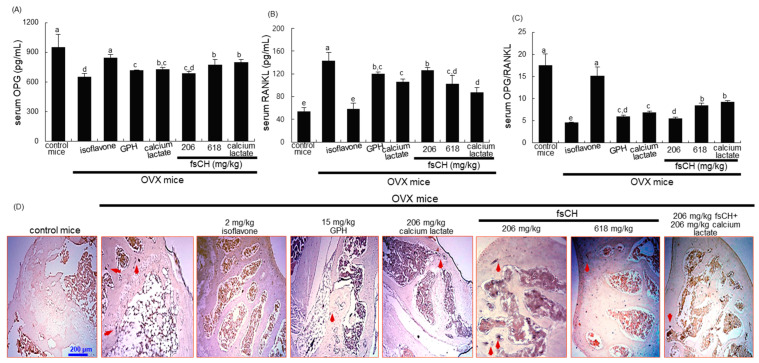
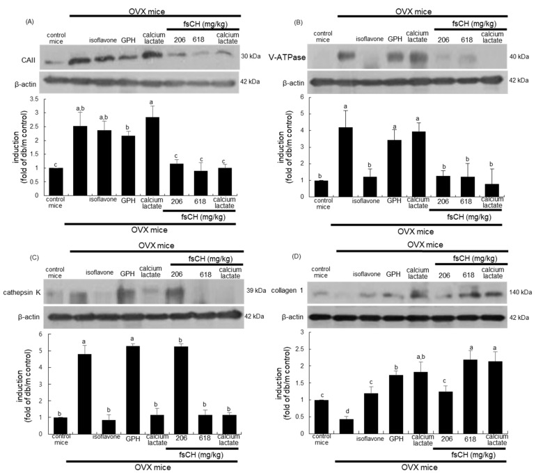
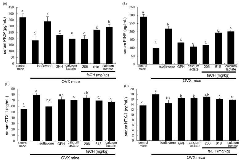
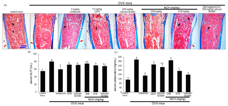
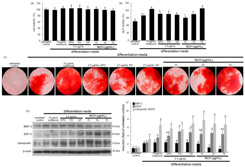
Osteoporosis manifest in postmenopausal women is an osteolytic disease characterized by bone loss, leading to increased susceptibility to bone fractures and frailty. The use of complementary therapies to alleviate postmenopausal osteoporosis is fairly widespread among women. The current study examined that Pangasius hypophthalmus fish skin collagen hydrolysates (fsCH) inhibited ovariectomy (OVX)-induced bone loss by conducting inter-comparative experiments for anti-osteoporotic activity among 206-618 mg/kg fsCH, 2 mg/kg isoflavone, 15 mg/kg glycine-proline-hydroxyproline (GPH) tripeptide, and calcium lactate. Surgical estrogen loss of mice for 8 weeks reduced serum 17β-estradiol levels with uterus atrophy, which was ameliorated by orally administering fsCH or isoflavone to mice. Similar to isoflavone, fsCH containing GPH-enhanced bone mineral density reduced levels of cathepsin K and proton-handling proteins, and elevated collagen 1 level in OVX bones. The treatment with fsCH and isoflavone enhanced the serum levels of collagen synthesis-related procollagen type 1 carboxy/amino-terminal propeptides reduced by OVX, whereas serum levels of osteocalcin and alkaline phosphatase, as well as collagen breakdown-related carboxy/amino-terminal telopeptides of type 1 collagen were reduced in OVX mice treated with fsCH, isoflavone, and calcium lactate. The trabecular bones were newly formed in OVX bones treated with isoflavone and fsCH, but not with calcium lactate. However, a low-dose combination of fsCH and calcium lactate had a beneficial synergy effect on postmenopausal osteoporosis. Furthermore, similar to isoflavone, 15-70 μg/mL fsCH, with its constituents of GPH and dipeptides of glycine-proline and proline-hydroxyproline, enhanced osteogenesis through stimulating differentiation, matrix mineralization, and calcium deposition of MC3T3-E1 osteoblasts. Accordingly, the presence of fsCH may encumber estrogen deficiency-induced bone loss through enhancing osteoclastogenic differentiation and matrix collagen synthesis. Therefore, fsCH may be a natural compound retarding postmenopausal osteoporosis and pathological osteoresorptive disorders.

Keywords: Pangasius hypophthalmus fish skin collagen hydrolysate; collagen; glycine–proline–hydroxyproline tripeptide; osteoblasts; ovariectomy; postmenopausal osteoporosis.


Conflict of interest statement

The authors declare no conflict of interest.

Figures

Figure 1
Figure 1
Uterus transverse section (A), wet weight of uterine tissues (B), and serum 17β-estradiol level (C) of ovariectomized (OVX) mice treated with various compounds daily for 8 weeks. OVX C57BL/6 female mice were orally administrated with 2 mg/kg isoflavone, 15 mg/kg glycine–proline–hydroxyproline tripeptide (GPH), 206 mg/kg calcium lactate, and 206–618 mg/kg Pangasius hypophthalmus fish skin hydrolysates (fsCH) daily for 8 weeks. Cross-sectional images of the uterine horn were obtained by staining with H&E, and visualized under light microscopy (A). Scale bar = 200 μm. Serum 17β-estradiol level was determined by using an ELISA kit (C). Respective values in bar graphs (mean ± SEM, n = 9) not having same alphabetical lowercase (a–e) are different at p < 0.05.
Figure 2
Figure 2
Effects of Pangasius hypophthalmus fish skin hydrolysates (fsCH) on serum osteoprotegerin (OPG)/receptor activator of the nuclear factors κB ligand (RANKL) ratio (AC), and TRAP localization in femoral bone tissue sections of OVX mice (D). Ovariectomized (OVX) C57BL/6 female mice were orally administrated with 2 mg/kg isoflavone, 15 mg/kg glycine–proline–hydroxyproline tripeptide (GPH), 206 mg/kg calcium lactate, and 206–618 mg/kg Pangasius hypophthalmus fish skin hydrolysates (fsCH) daily for 8 weeks. Serum levels of OPG and RANKL were determined by using ELISA kits (AC). Respective values in bar graphs (mean ± SEM, n = 9) not sharing an alphabetical lowercase (a–d) are different at p < 0.05. The TRAP staining of longitudinal femoral bone tissues was conducted by using a leukocyte acid phosphatase kit (D). TRAP-positive osteoclasts (purple) are observed at the femoral trabeculae. Scale bar = 200 μm. Representative images were visualized under light microscopy.
Figure 3
Figure 3
Inhibition of induction of carbonic anhydrase II (CAII, A), vacuolar-type H(+)- ATPase (V-ATPase, B), and cathepsin K (C), and increase in induction of collagen 1 (D) by Pangasius hypophthalmus fish skin hydrolysates (fsCH). OVX C57BL/6 female mice were orally administrated with 2 mg/kg isoflavone, 15 mg/kg glycine–proline–hydroxyproline tripeptide (GPH), 206 mg/kg calcium lactate, and 206–618 mg/kg fsCH daily for 8 weeks. Whole bone tissue extracts were subject to SDS-PAGE and Western blot analysis, with a specific antibody against CAII, V-ATPase, cathepsin K, and collagen 1. β-Actin was used as internal control. The bar graphs (mean ± SEM, n = 3) represent quantitative results of bands obtained from a densitometer. Values in respective bar graphs not having same alphabetical lowercase (a–d) are different at p < 0.05.
Figure 4
Figure 4
Effects of Pangasius hypophthalmus fish skin hydrolysates (fsCH) on collagen synthesis and degradation in ovariectomized (OVX) mice. OVX C57BL/6 female mice were orally administrated with 2 mg/kg isoflavone, 15 mg/kg glycine–proline–hydroxyproline tripeptide (GPH), 206 mg/kg calcium lactate, and 206–618 mg/kg Pangasius hypophthalmus fish skin hydrolysates (fsCH) daily for 8 weeks. Serum levels of procollagen type 1 carboxy-terminal propeptide (PICP, A), procollagen type 1 amino-terminal propeptide (PINP, B), carboxy-terminal telopeptide of type 1 collagen (CTX-1, C), and amino-terminal telopeptide of type 1 collagen (NTX-1, D) were measured by using ELISA kits. Respective values (mean ± SEM, n = 3) in bar graphs not sharing an alphabetical lowercase (a, b, c) are different at p < 0.05.
Figure 5
Figure 5
Inhibition of trabecular bone loss by Pangasius hypophthalmus fish skin hydrolysates (fsCH), and alterations in serum levels of alkaline phosphatase (ALP) and osteocalcin. OVX C57BL/6 female mice were orally administrated with 2 mg/kg isoflavone, 15 mg/kg glycine–proline–hydroxyproline tripeptide (GPH), 206 mg/kg calcium lactate, and 206–618 mg/kg fsCH daily for 8 weeks. Decalcified femoral bones of ovariectomized (OVX) mice were H&E-stained (A). Pink-stained mineralized bone, cortical bone outside, columns of trabecular bone inside. Scale bar = 100 μm. The serum level of ALP was measured by incubating with p-nitrophenyl phosphate and MgCl2 in Tris-HCl buffer. The absorbance was read at λ = 405 nm (B). The serum level of osteocalcin was measured by using an ELISA kit (C). Respective values (mean ± SEM, n = 3) in bar graphs not sharing an alphabetical lowercase (a-e) are different at p < 0.05.
Figure 6
Figure 6
MC3T3-E1 cell toxicity of Pangasius hypophthalmus fish skin hydrolysates (fsCH, A), upregulation of alkaline phosphatase (ALP) activity (B), calcium nodule formation by fsCH (C), and induction of non-collagenous matrix proteins (D). MC3T3-E1 cells were cultured for up to 21 days with 10 μg/mL isoflavone, 2.5 μg/mL glycine–proline–hydroxyproline tripeptide (GPH), 2.5 μg/mL proline–hydroxyproline dipeptide (PH), 2.5 μg/mL glycine–proline dipeptide (GP), and 15–70 μg/mL fsCH in differentiation media. Cell viability was measured by MTT assay (A). Bar graphs for viability (mean ± SEM, n = 3) was expressed as percentage of cell survival compared to untreated cells. MC3T3-E1 cells were cultured in differentiation media in the absence or presence of isoflavone, GPH, PH, GP, and fsCH for seven days. The ALP activity (B, mean ± SEM, n = 6) was measured at λ = 405 nm. Matrix mineralization was measured by Alizarin red S staining (C). Microphotographs were representative of 21 day-grown osteoblasts on the wells. Heavy reddish staining of Alizarin red S is proportional to the area of mineralized matrix in osteoblastic MC3T3-E1 cells. The calcium nodules were visualized under light microscopy (5 separate experiments). Magnification: 40-fold. Whole cell lysates were subject to SDS-PAGE and Western blot analysis with a specific antibody against bone morphogenetic protein-2 (BMP-2), bone sialoprotein II (BSPII), and osteopontin (D). β-Actin was used as internal control. The bar graphs (mean ± SEM, n = 3) represent quantitative results of bands obtained from a densitometer. Values in respective bar graphs not having same alphabetical lowercase (a–d) are different at p < 0.05.
Figure 7
Figure 7
Schematic diagram showing effects of Pangasius hypophthalmus fish skin hydrolysates (fsCH) on bone loss and osteoblastogenesis in ovariectomized mouse model. The arrows indicate activation by fsCH. ALP, alkaline phosphatase; BMD, bone mineral density; BSPII, bone sialoprotein II; CAII, carbonic anhydrase II; CTX-1, carboxy-terminal telopeptide of type 1 collagen; GPH, glycine–proline–hydroxyproline; NTX-1; amino-terminal telopeptide of type 1 collagen; OPG, osteoprotegerin; PICP, procollagen type 1 carboxy-terminal propeptide; PINP, procollagen type 1 amino-terminal propeptide; TRAP, tartrate-resistant acid phosphatase; RANK, receptor activator of nuclear factor-κB; RANKL, RANK ligand; V-ATPase, vacuolar-type H(+)- ATPase. The symbol → indicates activation manifested by fsCH and the symbol ⊗ indicates sites of inhibition.

Similar articles

Cited by

References

    1. Sözen T., Özışık L., Başaran N.Ç. An overview and management of osteoporosis. Eur. J. Rheumatol. 2017;4:46–56. doi: 10.5152/eurjrheum.2016.048. - DOI - PMC - PubMed
    1. Joestl J., Lang N., Bukaty A., Tiefenboeck T.M., Platzer P. Osteoporosis associated vertebral fractures-health economic implications. PLoS ONE. 2017;12:e0178209. doi: 10.1371/journal.pone.0178209. - DOI - PMC - PubMed
    1. Ji M.X., Yu Q. Primary osteoporosis in postmenopausal women. Chronic Dis. Transl. Med. 2015;1:9–13. - PMC - PubMed
    1. Eastell R., O’Neill T.W., Hofbauer L.C., Langdahl B., Reid I.R., Gold D.T., Cummings S.R. Postmenopausal osteoporosis. Nat. Rev. Dis. Primers. 2016;2:16069. doi: 10.1038/nrdp.2016.69. - DOI - PubMed
    1. Qadir A., Liang S., Wu Z., Chen Z., Hu L., Qian A. Senile osteoporosis: The involvement of differentiation and senescence of bone marrow stromal cells. Int. J. Mol. Sci. 2020;21:349. doi: 10.3390/ijms21010349. - DOI - PMC - PubMed

LinkOut - more resources