. 2022 Jun 11;10(6):1385.
doi: 10.3390/biomedicines10061385.

Macrophages Characterization in an Injured Bone Tissue

Affiliations

Macrophages Characterization in an Injured Bone Tissue

Krisztina Nikovics et al. Biomedicines. .

Abstract

Biomaterial use is a promising approach to facilitate wound healing of the bone tissue. Biomaterials induce the formation of membrane capsules and the recruitment of different types of macrophages. Macrophages are immune cells that produce diverse combinations of cytokines playing an important role in bone healing and regeneration, but the exact mechanism remains to be studied. Our work aimed to identify in vivo macrophages in the Masquelet induced membrane in a rat model. Most of the macrophages in the damaged area were M2-like, with smaller numbers of M1-like macrophages. In addition, high expression of IL-1β and IL-6 cytokines were detected in the membrane region by RT-qPCR. Using an innovative combination of two hybridization techniques (in situ hybridization and in situ hybridization chain reaction (in situ HCR)), M2b-like macrophages were identified for the first time in cryosections of non-decalcified bone. Our work has also demonstrated that microspectroscopical analysis is essential for macrophage characterization, as it allows the discrimination of fluorescence and autofluorescence. Finally, this work has revealed the limitations of immunolabelling and the potential of in situ HCR to provide valuable information for in vivo characterization of macrophages.

Keywords: Masquelet induced membrane; bone; cryosection; cytokines; hybridization chain reaction (HCR); macrophages.


Conflict of interest statement

The authors declare no conflict of interest.

Figures

Figure 1
Figure 1
Macrophage polarization subtypes; different cytokine expressions and functions of the macrophage populations in vitro and in vivo.
Figure 2
Figure 2
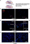
Histologic and microscopic analysis of rat femur: (AC) histologic analysis of the femurs; histopathological image of (A) non-operated; and (B) operated femurs of rats stained with hematoxylin and phloxin; (C) expanded view: high magnification image of the area within the red rectangle in image (B); (D,E) identification of macrophages M1 and M2 in the operated femur; anti-CD68 (Alexa488, turquoise fluorescence), labeling the M1 and M2 macrophages; anti-CD206 (Alexa568, red fluorescence) labeling the M2 macrophages and satellite cells; nuclear staining with DAPI (blue fluorescence).
Figure 3
Figure 3
Identification of M1-like and M2-like macrophages in the rat femurs: (A,B) immunolabeling with anti-CD68 and CD206 antibodies of the non-operated; and (CG) operated femurs; bright-field image together with immunolabeling of the non-operated (A) and operated (C) femurs; (EG) expanded view: high magnification image of the area within the red rectangle in image C; anti-CD68 (Alexa488, turquoise fluorescence), labeling the M1 and M2 macrophages; anti-CD206 (Alexa568, red fluorescence) labeling the M2 macrophages and satellite cells; nuclear staining with DAPI (blue fluorescence); thin arrow: M1-like macrophages; thick arrow: M2-like macrophages.
Figure 4
Figure 4
Identification of M2-like macrophages in the operated femur: (A) immunolabeling of the operated femur with anti-CD163; (B) negative control of the immunolabeling; anti-CD163 (Alexa488, turquoise fluorescence), labeling the M2 macrophages; nuclear staining with DAPI (blue fluorescence).
Figure 5
Figure 5
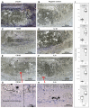
Identification of M1-like and M2-like macrophages in the rat femurs: (A) presentation of the different regions of the femurs; (B,D,F) operated; and (C,E,G) non-operated femurs labeled with anti-CD68 and anti-CD206 antibodies; anti-CD68 (Alexa488, turquoise fluorescence), labeling the M1 and M2 macrophages; anti-CD206 (Alexa568, red fluorescence) labeling the M2 macrophages and satellite cells; nuclear staining with DAPI (blue fluorescence); thin arrow: satellite cells; arrowhead: osteoclast.
Figure 6
Figure 6
Observation of the autofluorescence in the operated rat femur: (A) detected autofluorescence after excitation with light (488 wavelengths, turquoise fluorescence); (B) detected autofluorescence after excitation with light (568 wavelengths, red fluorescence); (C) nuclear staining with DAPI (blue fluorescence); (D) merged image; (E) bright-field image; (F) expanded view: high magnification image of the area within the red rectangle in image (D); (G) expanded view: high magnification image of the area within the red rectangle in image (E).
Figure 7
Figure 7
Identification of the in situ autofluorescence by microspectroscopy analysis. In the operated femur, macrophages were labeled with anti-CD68 antibody. Tissues were excited at 488 nm. (A) Emission of the autofluorescence and the antigen-specific fluorescence (Alexa Fluor 488) with turquoise fluorescence. (B) Microspectroscopycal analysis of Alexa Fluor 488 emission (turquoise line) and autofluorescence (red line). (C) Separation of autofluorescence emission (red fluorescence) and antigen-specific fluorescence (Alexa Fluor 488) (turquoise fluorescence). (D) Emission of the autofluorescence (red fluorescence). (E) Emission of the antigen-specific fluorescence (Alexa Fluor 488) (turquoise fluorescence). Nuclear staining with DAPI (blue fluorescence). Fine arrow: macrophages.
Figure 8
Figure 8
Expression of the different marker genes and cytokines in the operated femur; qRT-PCR analysis of the different gene expressions. This result is the average of three measurements.
Figure 9
Figure 9
In situ hybridization in the operated rat femur: (A) expression of β-actin mRNA (positive control); (B) negative control; (C) CD68 mRNA; (D) IL-1β mRNA; (E) CD163 mRNA; (F) IL-6 mRNA; (G) expanded view: high magnification image of the area within the red rectangle in image (E); (H) expanded view: high magnification image of the area within the red rectangle in image (F); (I) the stained area of the picture was quantitatively analyzed using ImageJ. The Masquelet induced membrane region was compared with the control region (C) (n = 3). ** p < 0.01, * p < 0.5, ns = not significant, compared to control region.
Figure 10
Figure 10
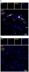
Identification of the M2b-like macrophages in the operated femur by in situ hybridization combined with hybridization-chain-reaction detection (in situ HCR): (A) in situ hybridization combined with hybridization-chain-reaction detection (in situ HCR); (B) negative control: Probe-IL-6 (Alexa488, turquoise fluorescence), Probe-IL-1β (Alexa546, red fluorescence), Probe-CD163 (Alexa647, yellow fluorescence), nuclear staining with DAPI (blue fluorescence).

References

    1. Pajarinen J., Lin T., Gibon E., Kohno Y., Maruyama M., Nathan K., Lu L., Yao Z., Goodman S.B. Mesenchymal Stem Cell-Macrophage Crosstalk and Bone Healing. Biomaterials. 2019;196:80–89. doi: 10.1016/j.biomaterials.2017.12.025. - DOI - PMC - PubMed
    1. Sun Y., Li J., Xie X., Gu F., Sui Z., Zhang K., Yu T. Macrophage-Osteoclast Associations: Origin, Polarization, and Subgroups. Front. Immunol. 2021;12:778078. doi: 10.3389/fimmu.2021.778078. - DOI - PMC - PubMed
    1. Smith J.K. Osteoclasts and Microgravity. Life. 2020;10:207. doi: 10.3390/life10090207. - DOI - PMC - PubMed
    1. Gibon E., Lu L., Goodman S.B. Aging, Inflammation, Stem Cells, and Bone Healing. Stem. Cell Res. Ther. 2016;7:44. doi: 10.1186/s13287-016-0300-9. - DOI - PMC - PubMed
    1. Weitzmann M.N. Bone and the Immune System. Toxicol. Pathol. 2017;45:911–924. doi: 10.1177/0192623317735316. - DOI - PMC - PubMed

LinkOut - more resources