. 2022 Jun 13;10(6):1397.
doi: 10.3390/biomedicines10061397.

Murine Falcor/LL35 lncRNA Contributes to Glucose and Lipid Metabolism In Vitro and In Vivo

Affiliations

Murine Falcor/LL35 lncRNA Contributes to Glucose and Lipid Metabolism In Vitro and In Vivo

Evgeniya Shcherbinina et al. Biomedicines. .

Abstract

Glucose and lipid metabolism are crucial functional systems in eukaryotes. A large number of experimental studies both in animal models and humans have shown that long non-coding RNAs (lncRNAs) play an important role in glucose and lipid metabolism. Previously, human lncRNA DEANR1/linc00261 was described as a tumor suppressor that regulates a variety of biological processes such as cell proliferation, apoptosis, glucose metabolism and tumorigenesis. Here we report that murine lncRNA Falcor/LL35, a proposed functional analog of human DEANR1/linc00261, is predominantly expressed in murine normal hepatocytes and downregulated in HCC and after partial hepatectomy. The application of high-throughput approaches such as RNA-seq, LC-MS proteomics, lipidomics and metabolomics analysis allowed changes to be found in the transcriptome, proteome, lipidome and metabolome of hepatocytes after LL35 depletion. We revealed that LL35 is involved in the regulation of glycolysis and lipid biosynthesis in vitro and in vivo. Moreover, LL35 affects Notch and NF-κB signaling pathways in normal hepatocytes. All observed changes result in the decrease in the proliferation and migration of hepatocytes. We demonstrated similar phenotype changes between murine LL35 and human linc00261 depletion in vitro and in vivo that opens the opportunity to translate results for LL35 from a liver murine model to possible functions of human lncRNA linc00261.

Keywords: glucose metabolism; hepatocyte; lipid metabolism; liver; long non-coding RNA.


Conflict of interest statement

The authors declare no conflict of interest.

Figures

Figure 1
Figure 1
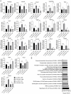
Evaluation of LL35 RNA levels in vitro and in vivo by RT-qPCR. (A) Hepatocyte cell lines. (B) Normal and HCC mouse liver. (C) Partial hepatectomy. (D) LL35 KD in the liver at day 5 after injection of GalNac-ASO conjugates (LL35 ASO). Luc ASO—control. ACTB—reference gene in RT-qPCR. Results show mean ± SD. * p < 0.05, ** p < 0.01 and *** p < 0.001.
Figure 2
Figure 2
Results of transcriptome analysis of AML12 cells and murine liver after LL35 depletion. (A) Venn diagram for differentially expressed genes after LL35 knockdown in AML12 cells in comparison with murine liver. Top representative pathways with p-value < 0.05 obtained by PANTHER Reactome analysis of differentially expressed genes after LL35 depletion in (B) AML12 cells; (C) murine liver.
Figure 3
Figure 3
Transcriptome validation and proteome analysis of AML12 cells and murine liver after LL35 depletion. (A) Validation of transcriptome data by RT-qPCR, normalization on ACTB gene. (B) Top representative pathways obtained by PANTHER Reactome analysis for significantly changed proteins after LL35 depletion in AML12 cells. Results show mean ± SD. ns—not significant. * p < 0.05, ** p < 0.01 and *** p < 0.001.
Figure 4
Figure 4
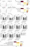
Lipidome and metabolome analysis of AML12 cells and murine liver after LL35 inhibition. (A) PCA of lipids and metabolites from AML12 cells and liver samples with depleted LL35 (LL35 ASO) and control (Luc ASO), polarities merged. Lipid abundance alteration in (B) AML12 cells; (C) murine liver, classes that are significant both before and after confirmation (fgsea FDR < 10%). Numbers in brackets: the number of confirmed features and the total number of features. (D) RT-qPCR analysis of expression levels of key genes, which participate in synthesis and breakdown of significantly changed lipids classes after LL35 knockdown (LL35 ASO) and control (Luc ASO), normalization on ACTB gene. (E) KEGG pathways, metabolites from which are significantly influenced by LL35 knockdown in AML12 cells. NES—normalized enrichment score. Results show mean ± SD. ns—not significant. * p < 0.05, ** p < 0.01 and *** p < 0.001.
Figure 5
Figure 5
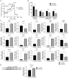
Changes in the glucose metabolism after LL35 knockdown in vitro. (A) An averaged time curve for ECAR after subsequent injections of 10 mM glucose, 1 μM oligomycin and 50 mM 2-DG. Each data point represents an ECAR value used for calculations. (B) Individual parameters for cell glycolytic function, including glycolytic capacity, glycolysis, non-glycolytic acidification and glycolytic reserve, for AML12 cells after LL35 depletion (LL35 ASO) and control (Luc ASO). (C) Evaluation of mRNA levels for key genes and transcription factors, which participate in glucose metabolism after LL35 knockdown (LL35 ASO) and control (Luc ASO) by RT-qPCR with normalization on ACTB gene. (D) Western blot and its quantification of PEPCK protein levels in AML12 cells after LL35 inhibition (LL35 ASO) and control (Luc ASO). Results show mean ± SD. ns—not significant. * p < 0.05, ** p < 0.01 and *** p < 0.001.
Figure 6
Figure 6
The influence of insulin in mediating the effects of LL35 depletion. (A) Insulin tolerance test in vivo. Glucose blood level in mice was measured each 15 min for 1 h after insulin injection (1 U/kg). LL35 ASO—mice with depleted LL35, Luc ASO—control mice. (B) Biochemical parameters of mice blood at day 5 after LL35 depletion (LL35 ASO) in comparison to control mice (Luc ASO). Insulin treatment compensates these differences. (C) Western blot of pAKT1 and AKT1 proteins in AML12 cells after LL35 knockdown (LL35 ASO) and control (Luc ASO) with 100 nM (+Ins) or without insulin treatment. (D) Quantification of pAKT1/AKT1 ratio for AML12 cells after LL35 knockdown and insulin treatment. Results show mean ± SD. ns—not significant. * p < 0.05, ** p < 0.01.
Figure 7
Figure 7
(A) Study of hepatocyte survival after LL35 depletion (LL35 ASO) in comparison with control cells (Luc ASO). (B) Wound healing assay for AML12 cells after LL35 knockdown and control. (C) Quantification of wound healing assay with ImageJ software. (D) Cell cycle analysis of the cells with LL35 knockdown and control cells by flow cytometry. Percentage of cells in different cell cycle phases after LL35 knockdown compared with control obtained from flow cytometry analysis after PI staining. Results show mean ± SD. ns—not significant. * p < 0.05, ** p < 0.01 and *** p < 0.001.
Figure 8
Figure 8
Changes in Notch and NF-κB pathways after LL35 depletion in vitro. (A) Estimation of Jagged1 protein level after LL35 knockdown in vitro by western blot, normalized at ActB protein level. (B) RT-qPCR measurement of the mRNA expression levels of major genes involved in Notch signaling pathway after LL35 knockdown (LL35 ASO) in AML12 cells and control (Luc ASO), normalization on ACTB gene. (C) Estimation of p105/p50 proteins’ ratio after LL35 knockdown in AML12 cells by western blot, normalized on ActB protein level. (D) Estimation of IκBa protein level after LL35 knockdown by western blot, normalization on GAPDH protein level. (E) RT-qPCR measured expression levels of IκBa mRNA after LL35 knockdown and control. ACTB—reference gene. Results show mean ± SD. ns—not significant. * p < 0.05, ** p < 0.01.

Similar articles

References

    1. Djebali S., Davis C.A., Merkel A., Dobin A., Lassmann T., Mortazavi A.M., Tanzer A., Lagarde J., Lin W., Schlesinger F., et al. Landscape of Transcription in Human Cells. Nature. 2012;489:101–108. doi: 10.1038/nature11233. - DOI - PMC - PubMed
    1. Moore J.B., Uchida S. Functional Characterization of Long Non-Coding RNAs. Curr. Opin. Cardiol. 2020;35:199–206. doi: 10.1097/HCO.0000000000000725. - DOI - PMC - PubMed
    1. Bhat S.A., Ahmad S.M., Mumtaz P.T., Malik A.A., Dar M.A., Urwat U., Shah R.A., Ganai N.A. Long Non-Coding RNAs: Mechanism of Action and Functional Utility. Non-Coding RNA Res. 2016;1:43–50. doi: 10.1016/j.ncrna.2016.11.002. - DOI - PMC - PubMed
    1. Huang B., Zhang R. Regulatory Non-Coding RNAs: Revolutionizing the RNA World. Mol. Biol. Rep. 2014;41:3915–3923. doi: 10.1007/s11033-014-3259-6. - DOI - PubMed
    1. Lin Y.-H. Crosstalk of LncRNA and Cellular Metabolism and Their Regulatory Mechanism in Cancer. Int. J. Mol. Sci. 2020;21:2947. doi: 10.3390/ijms21082947. - DOI - PMC - PubMed