Review
. 2022 Jun 17;10(6):1432.
doi: 10.3390/biomedicines10061432.

Molecular Pathophysiological Mechanisms in Huntington's Disease

Affiliations
Review

Molecular Pathophysiological Mechanisms in Huntington's Disease

Anamaria Jurcau. Biomedicines. .

Abstract

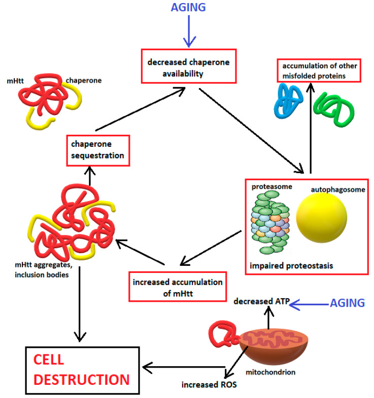
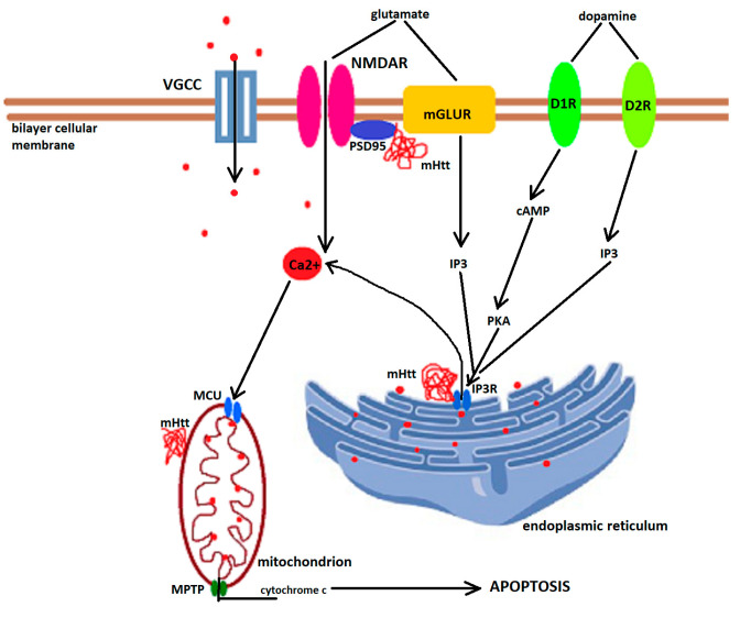
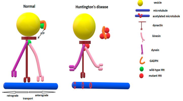
Huntington's disease is an inherited neurodegenerative disease described 150 years ago by George Huntington. The genetic defect was identified in 1993 to be an expanded CAG repeat on exon 1 of the huntingtin gene located on chromosome 4. In the following almost 30 years, a considerable amount of research, using mainly animal models or in vitro experiments, has tried to unravel the complex molecular cascades through which the transcription of the mutant protein leads to neuronal loss, especially in the medium spiny neurons of the striatum, and identified excitotoxicity, transcriptional dysregulation, mitochondrial dysfunction, oxidative stress, impaired proteostasis, altered axonal trafficking and reduced availability of trophic factors to be crucial contributors. This review discusses the pathogenic cascades described in the literature through which mutant huntingtin leads to neuronal demise. However, due to the ubiquitous presence of huntingtin, astrocytes are also dysfunctional, and neuroinflammation may additionally contribute to Huntington's disease pathology. The quest for therapies to delay the onset and reduce the rate of Huntington's disease progression is ongoing, but is based on findings from basic research.

Keywords: Huntington’s disease; animal models; brain-derived neurotrophic factor; excitotoxicity; mitochondrial dysfunction; mutant huntingtin; oxidative stress; proteostasis; transcriptional dysregulation.


Conflict of interest statement

The author declares no conflict of interest.

Figures

Figure 1
Figure 1
Impaired proteostasis in Huntington’s disease. The chronic production of misfolded huntingtin protein acts synergistically with aging to decrease available chaperones. This leads to accumulation of other misfolded proteins. The mHtt eventually overwhelms the proteasome and impairs the misfolded protein clearance system, an impairment further augmented by the limited availability of ATP due to aging and mHtt-related mitochondrial dysfunction leading to increases in the production of reactive oxygen species (ROS), which, in turn, potentiate mitochondrial dysfunction in a vicious cascade. The accumulation of mHtt promotes its aggregation, further sequestering chaperones in a vicious cycle that disrupts cellular homeostasis and culminates in cell destruction.
Figure 2
Figure 2
Impaired calcium homeostasis in Huntington’s disease leading to apoptosis. Mutant huntingtin (mHtt) enhances NMDA receptor (NMDAR) function, possibly through decreased interaction with the PDS95 (postsynaptic density 95)/NMDAR complex. Dopamine released by the dopaminergic neurons acts on D1 receptors (D1R), which activate adenyl cyclase, the increased cAMP levels activating phosphokinase A (PKA), which, in turn, stimulates the inositol 1,4,5-phosphate receptors (IP3R), while dopamine acting on D2 receptors directly activate IP3R. IP3R activation leads to release of Ca2+ from the endoplasmic reticulum. Excessive cytosolic calcium is taken up by mitochondria through the mitochondrial calcium uniporter (MCU), but will lead to opening of the mitochondrial permeability transition pore (MPTP), especially when mHtt directly associates with mitochondrial membrane proteins. VGCC—L-type voltage-gated calcium channel; red dots—calcium; red whirl—aberrantly folded mutant huntingtin protein.
Figure 3
Figure 3
The involvement of Htt in normal vesicular transport and role of mHtt in impaired vesicular transport in HD. Under normal conditions, HTT interacts with the motor proteins dynactin, dynein, and kinesin and recruits GADPH to supply the necessary ATP. In HD, mHtt aggregates sequester GAPDH and motor proteins, and phosphorylates kinesin. Furthermore, microtubules are acetylated by mHtt, which hinders binding of kinesin-1 to the microtubules.

References

    1. Zuccato C., Valenza M., Cattaneo E. Molecular mechanisms and potential therapeutic targets in Huntington’s disease. Physiol. Rev. 2010;90:905–981. doi: 10.1152/physrev.00041.2009. - DOI - PubMed
    1. Perandones C., Radrizzani M., Micheli F.E. Molecular Mechanisms Involved in the Pathogenesis of Huntington’s Disease. In: Visser T.J., editor. Huntington’s Disease: Etiology and Symptoms, Diagnosis and Treatment. Nova Science Publishers, Inc.; New York, NY, USA: 2010. pp. 1–37.
    1. Gusella J.F., Wexler N.S., Conneally P.M., Naylor S.L., Anderson M.A., Tanzi R.E., Watkins P.C., Ottina K., Wallace M.R., Sakaguchi A.Y., et al. A polymorphic DNA marker genetically linked to Huntington’s disease. Nature. 1983;306:234–238. doi: 10.1038/306234a0. - DOI - PubMed
    1. HD Collaborative Research Group A novel gene containing a trinucleotide repeat that is expanded and unstable on Huntington’s disease chromosomes. Cell. 1993;72:971–983. doi: 10.1016/0092-8674(93)90585-E. - DOI - PubMed
    1. Ranen N.G., Stine O.C., Abbott M.H., Sherr M., Codori A.M., Franz M.L., Chao N.I., Chung A.S., Pleasant N., Callahan C. Anticipation and instability of IT-15 (CAG)n repeats in parent-offspring pairs with Huntington’s disease. Am. J. Hum. Genet. 1995;57:593–602. - PMC - PubMed

LinkOut - more resources