Review
. 2022 Jun 8;14(12):2836.
doi: 10.3390/cancers14122836.

Non-Melanoma Skin Cancer Clearance after Medical Treatment Detected with Noninvasive Skin Imaging: A Systematic Review and Meta-Analysis

Affiliations
Review

Non-Melanoma Skin Cancer Clearance after Medical Treatment Detected with Noninvasive Skin Imaging: A Systematic Review and Meta-Analysis

Stefania Guida et al. Cancers (Basel). .

Abstract

Background/objectives: Non-melanoma skin cancer (NMSC) treated with nonsurgical therapies can be monitored with noninvasive skin imaging. The precision of dermoscopy, reflectance confocal microscopy (RCM) and optical coherence tomography (OCT) in detecting clearance is unclear. We aim to report the proportion of persisting tumors identified with noninvasive technologies available in the literature.

Methods: A systematic literature search was conducted on the PubMed and Cochrane Public Library Databases for articles published prior to November 2021. Statistical analyses were conducted with MedCalc 14.8.1 software.

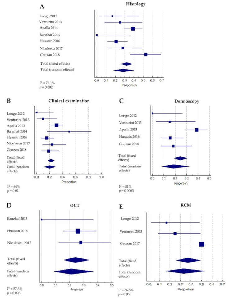
Results: A total of eight studies (352 lesions) reporting noninvasive imaging for NMSC clearance following nonsurgical treatment were included. Most (n = 7) reported basal cell carcinoma (BCC), and one study reported squamous cell carcinoma (SCC) clearance. A meta-analysis of the BCC clearance revealed that the summary effect for RCM was higher, as compared to the other techniques. Interestingly, the sensitivity and specificity for OCT were 86.4% (95% CI: 65.1-97.1) and 100% (95% CI: 94.8-100.0), respectively, whilst, for RCM, they reached 100% (95%CI: 86.8-100) and 72.5% (95% CI: 64.4-79.7), respectively.

Conclusions: Routine clinical examination and dermoscopy underperform when employed for NMSC clearance monitoring, although they represent the first approach to the patient. OCT and RCM seem to improve the detection of persistent BCC after medical treatment.

Keywords: basal cell carcinoma; dermoscopy; non-melanoma skin cancer; nonsurgical treatment; optical coherence tomography; reflectance confocal microscopy; squamous cell carcinoma.


Conflict of interest statement

The authors declare no conflict of interest.

Figures

Figure 1
Figure 1
Literature flow diagram for the search and study selections.
Figure 2
Figure 2
Proportion of the included studies with different detection methods and rates of persistent basal cell carcinomas.
Figure 3
Figure 3
Proportion of included studies and rates of persistent tumors with (A) histology, (B) clinical examination, (C) dermoscopy, (D) optical coherence tomography, (OCT) and (E) reflectance confocal microscopy (RCM) [15,29,30,31,32,33,34].
Figure 4
Figure 4
(A) Clinical image of a superficial basal cell carcinoma (BCC) from our center archive located on the left forehead of a 60-year-old woman, before initiating imiquimod 5% cream applied 5 times a week for 6 weeks. (B) Clinical image two months after the six-week cycle of treatment showing erythema, crusting and erosion. (C) Complete clinical healing of the lesion at the 18-month follow-up visit was observed. (D) Dermoscopic image of the superficial basal cell carcinoma (BCC) showing small erosions at the baseline and (E) after imiquimod treatment. Dermoscopic clearance was observed at the 18-month follow-up. (F) Corresponding OCT image shows ovoid structures protruding from the epidermis (white rectangular area) at the baseline and (G) complete OCT clearance at the 18-month follow-up (image size 6 mm × 2 mm). (H) RCM image shows tumor cords (white arrows) of the BCC at the baseline (I), which were no longer observed at the 18-month follow-up (scale bar 100 µm), confirming the RCM clearance.

References

    1. Conforti C., Corneli P., Harwood C., Zalaudek I. Evolving Role of Systemic Therapies in Non-Melanoma Skin Cancer. Clin. Oncol. (R. Coll. Radiol.) 2019;31:759–768. doi: 10.1016/j.clon.2019.08.011. - DOI - PubMed
    1. Rogers H.W., Weinstock M.A., Harris A.R., Hinckley M.R., Feldman S.R., Fleischer A.B., Coldiron B.M. Incidence Estimate of Nonmelanoma Skin Cancer in the United States, 2006. Arch. Dermatol. 2010;146:283–287. doi: 10.1001/archdermatol.2010.19. - DOI - PubMed
    1. Kauvar A.N.B., Cronin T., Roenigk R., Hruza G., Bennett R. Consensus for Nonmelanoma Skin Cancer Treatment: Basal Cell Carcinoma, Including a Cost Analysis of Treatment Methods. Dermatol. Surg. 2015;41:550–571. doi: 10.1097/DSS.0000000000000296. - DOI - PubMed
    1. Kauvar A.N.B., Arpey C.J., Hruza G., Olbricht S.M., Bennett R. Consensus for Nonmelanoma Skin Cancer Treatment, Part II: Squamous Cell Carcinoma, Including a Cost Analysis of Treatment Methods. Dermatol. Surg. 2015;41:1214–1240. doi: 10.1097/DSS.0000000000000478. - DOI - PubMed
    1. Mun J.-H., Park J.-M., Song M., Jwa S.-W., Kim H.-S., Ko H.-C., Kim B.-S., Kim M.-B. The Use of Dermatoscopy to Monitor Therapeutic Response of Bowen Disease: A Dermatoscopic Pathological Study. Br. J. Dermatol. 2012;167:1382–1385. doi: 10.1111/j.1365-2133.2012.11124.x. - DOI - PubMed