Review
. 2022 Jun 11;14(12):2885.
doi: 10.3390/cancers14122885.

Current and Emerging Methods for Ovarian Cancer Screening and Diagnostics: A Comprehensive Review

Affiliations
Review

Current and Emerging Methods for Ovarian Cancer Screening and Diagnostics: A Comprehensive Review

Juliane M Liberto et al. Cancers (Basel). .

Abstract

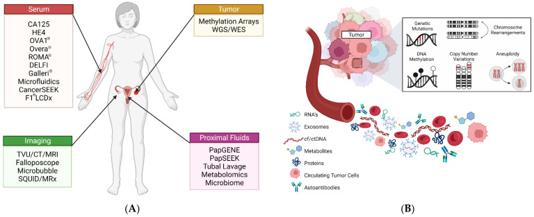
With a 5-year survival rate of less than 50%, ovarian high-grade serous carcinoma (HGSC) is one of the most highly aggressive gynecological malignancies affecting women today. The high mortality rate of HGSC is largely attributable to delays in diagnosis, as most patients remain undiagnosed until the late stages of -disease. There are currently no recommended screening tests for ovarian cancer and there thus remains an urgent need for new diagnostic methods, particularly those that can detect the disease at early stages when clinical intervention remains effective. While diagnostics for ovarian cancer share many of the same technical hurdles as for other cancer types, the low prevalence of the disease in the general population, coupled with a notable lack of sensitive and specific biomarkers, have made the development of a clinically useful screening strategy particularly challenging. Here, we present a detailed review of the overall landscape of ovarian cancer diagnostics, with emphasis on emerging methods that employ novel protein, genetic, epigenetic and imaging-based biomarkers and/or advanced diagnostic technologies for the noninvasive detection of HGSC, particularly in women at high risk due to germline mutations such as BRCA1/2. Lastly, we discuss the translational potential of these approaches for achieving a clinically implementable solution for screening and diagnostics of early-stage ovarian cancer as a means of ultimately improving patient outcomes in both the general and high-risk populations.

Keywords: HGSC; biomarkers; diagnostic; emerging; ovarian cancer; screening.


Conflict of interest statement

The authors declare no conflict of interest.

Figures

Figure 1
Figure 1
Overview of ovarian cancer screening and diagnostics. (A) Relevant emerging strategies for screening and detection of HGSC. (B) Circulating tumor biomarkers that can be assayed for screening and diagnostic purposes.
Figure 2
Figure 2
Representative sonographic images of benign (B1B5) and malignant (M1M5) ovarian masses. Features in each panel are reviewed based on the IOTA simple rules to evaluate malignancy of ovarian lesions. In general, the presence of irregular shaped bodies, papillary projections, and/or internal blood flow are predictive of malignancy. Images provided from Kaijser et al. [41]. Used with permission.
Figure 3
Figure 3
Molecular model and array construction of OCC-DNA nanosensor elements created by Kim et al. [138]. (A) Fabricated molecular model of a single OCC-DNA nanosensor with ssDNA wrapped around a semi-conducting single-walled nanotube (SWNCT) modified with organic color sensors (OCC). (B) Nanosensor array construction is generated from a matrix combination of OCC-modified SWNCTs with DNA. Used with permission.
Figure 4
Figure 4
Schematic of the antibody-lectin barcode microfluidic platform proposed by Shang et al. [140]. (A) The integrated microfluidic lectin barcode platform contains eight identical microchambers that lay perpendicular to an array of lectins patterned on the chip’s surface. Pneumatic actuation within the assay chamber allows for mixing and capture of glycoproteins to the lectin array. (B) Digital image of the microchip with loading chambers in red and pneumatic control layers in green. Reprinted under the Creative Commons CC BY-4.0 license.
Figure 5
Figure 5
Principles of contrast-enhanced ultrasound imaging with contrast microbubble reported by Pysz et al. [147] Microbubbles generally consist of a gaseous core that is enveloped in a lipid or protein shell that may or may not be “decorated” with additional components for stability. Due to their micron size, microbubbles are able to travel within tiny microvascular spaces, including microcapillaries that may be present in solid lesions. Used with permission.
Figure 6
Figure 6
Biomarkers used in the OVA1® and Overa® assays developed by Aspira Women’s Health Inc. (A) The OVA1® assay incorporates TVU findings and menopausal status with changes in serum proteins ApoA-1, B2M, CA125, Transthyretin (Prealbumin) and Transferrin. (B) The Overa® assay is the second-generation model which incorporates two new serum biomarkers HE4 and FSH, along with three of the original biomarkers defined in the OVA1® assay. Image was recreated with permission from Aspira Women’s Health [176].
Figure 7
Figure 7
PapSEEK, proposed by Wang et al. [90], (A) DNA extracted from patient cytologic fluids and plasma is assayed for genetic alterations. (B) Target genes assayed for mutations were found in cytologic fluid for endometrial cancer samples and in both cytologic fluids and plasma for ovarian cancer samples but not in healthy controls. Used with permission.
Figure 8
Figure 8
Schematic of DELFI approach. Cristiano et al. [233]. Cancer-specific fragment profiles are generated through WGS of cfDNA isolated from plasma and compared to profiles generated from healthy individuals. Unique fragment alterations at specific loci can be mapped back to detect and identify specific cancer types. Used with permission.
Figure 9
Figure 9
Workflow of the ExoSearch chip developed by Zhao et al. [257]. (A) Patient plasma (orange) is mixed with immunomagnetic beads that bind exosomes within the sample. Beads carrying exosomes are then isolated via a magnetized field in which the number of beads isolated was in direct comparison to the sample input and could be quantified. A mixture of fluorescently labeled antibodies is then applied to the isolated beads for multi-color fluorescence imaging. (B,C) Bright-field images of the immunomagnetic beads in the microfluidic compartments. (D) Aggregated exosome-bound immunomagnetic beads after magnetic separation (E) Transmission electron micrograph depicting the cross-section of an exosome-bound immunomagnetic bead. Reprinted under the Creative Commons CC BY-NC3.0 license.

References

    1. Shih I.M., Wang Y., Wang T.L. The Origin of Ovarian Cancer Species and Precancerous Landscape. Am. J. Pathol. 2021;191:26–39. doi: 10.1016/j.ajpath.2020.09.006. - DOI - PMC - PubMed
    1. Rendi M.H. Epithelial carcinoma of the ovary, fallopian tube, and peritoneum: Histopathology. In: Chakrabarti A., editor. UpToDate. UpToDate; Waltham, MA, USA: 2021.
    1. Bowtell D.D., Böhm S., Ahmed A.A., Aspuria P.J., Bast R.C., Beral V., Berek J.S., Birrer M.J., Blagden S., Bookman M.A., et al. Rethinking ovarian cancer II: Reducing mortality from high-grade serous ovarian cancer. Nat. Rev. Cancer. 2015;15:668–679. doi: 10.1038/nrc4019. - DOI - PMC - PubMed
    1. Siegel R.L., Miller K.D., Fuchs H.E., Jemal A. Cancer statistics, 2022. CA Cancer J. Clin. 2022;72:7–33. doi: 10.3322/caac.21708. - DOI - PubMed
    1. Berek J.S., Kehoe S.T., Kumar L., Friedlander M. Cancer of the ovary, fallopian tube, and peritoneum. Int. J. Gynecol. Obstet. 2018;143:59–78. doi: 10.1002/ijgo.12614. - DOI - PubMed

LinkOut - more resources