Review
. 2022 Jun 15;14(12):2954.
doi: 10.3390/cancers14122954.

A Tale of Two Cancers: A Current Concise Overview of Breast and Prostate Cancer

Affiliations
Review

A Tale of Two Cancers: A Current Concise Overview of Breast and Prostate Cancer

Franklyn De Silva et al. Cancers (Basel). .

Abstract

Cancer is a global issue, and it is expected to have a major impact on our continuing global health crisis. As populations age, we see an increased incidence in cancer rates, but considerable variation is observed in survival rates across different geographical regions and cancer types. Both breast and prostate cancer are leading causes of morbidity and mortality worldwide. Although cancer statistics indicate improvements in some areas of breast and prostate cancer prevention, diagnosis, and treatment, such statistics clearly convey the need for improvements in our understanding of the disease, risk factors, and interventions to improve life span and quality of life for all patients, and hopefully to effect a cure for people living in developed and developing countries. This concise review compiles the current information on statistics, pathophysiology, risk factors, and treatments associated with breast and prostate cancer.

Keywords: breast cancer; cancer classification; cancer diagnosis; cancer heterogeneity; cancer pathophysiology; cancer statistics; cancer treatment; drug-tolerant persister cells; female breast anatomy; multidrug resistance; neuroendocrine cancer; prostate anatomy; prostate cancer; risk factors.


Conflict of interest statement

The authors declare no conflict of interest.

Figures

Figure 1
Figure 1
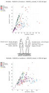
A snapshot of worldwide cancer incidence and mortality: Representation of the major cancer types (color coded) in each country based on the age standardized incidence rates for all ages and sex (world map). On the right, the bar graph represents the age standardized rates (ASR) based estimates on incidence and mortality, accounting for all ages for both male and female combined. Bottom left dot plot represents the ASR estimates on the world human development index (HDI) vs. incidence. Bottom right graph represents the global ASR estimates on the incidence vs mortality. The incidence, mortality, and prevalence information presented on ‘CANCER TODAY’ (World Health Organization) represents a collection of 36 specific types of cancer and 185 countries or territories of the world in 2020 (GLOBOCAN project, International Agency for Research on Cancer (IARC)), obtained from many cancer sites from across the globe. Figures taken from IARC website (information publicly available online accessed in 20 November 2021, https://gco.iarc.fr/today/home) [9,14,15].
Figure 2
Figure 2
2020 Global breast and prostate cancer statistics for women and men: Estimated number of new cases and deaths for both sexes including all ages. The top graph represents prostate cancer and bottom graph represents breast cancer: age standardized rates (ASR) based estimated new cases and deaths as reported by Global Cancer Observatory (GCO). Both breast and prostate cancer are among the leading cancers contributing to both new cases and deaths in women and men. Figures taken from IARC website (https://gco.iarc.fr/today, accessed on 20 November 2021) [10,14,15].
Figure 3
Figure 3
Schematic view of the human female breast and different types of interactive cells that are present within the breast tissue. The breast tissue overlays the ribs and chest muscles. The adult woman’s breast contains glandular epithelium (~10–15%) and this milk producing epithelia is contained within the surrounding adipose tissue. Multiple lobules (terminal ductules, acini, milk-producing lobules) together make up the lobes of the breast. The functional units of the breast are the terminal duct lobular units. All lobules and lobes are connected to the nipple through a branched system of ducts. Terminal ductal lobular units (TDLUs), which is a collection of ductules, intralobular duct, loose intralobular connective tissue, and extralobular terminal duct, are common sites of origin for several breast cancers. Within the stroma, two types of fibroblasts are present. Loosely connected intralobular fibroblasts surround the epithelial cells and they are subsequently encompassed by the more condensed interlobular fibroblasts. The other important cellular component of the mammary stroma is adipocytes (i.e., fat cells). The parenchymal tissue consists of epithelial and myoepithelial cells. In addition, the stromal compartment contains vascular endothelial cells and infiltrating immune cells. Stromal cells secrete factors of the extracellular matrix (e.g., collagens, hyaluronic acid, tenascins, fibronectins, proteoglycans) that are integral for the breast’s three-dimensional microstructure. Mammary ducts consist of polarized apically orientated columnar luminal epithelial cells that line (inside) ducts along with alveolar structures at the ends, as well as contractile myoepithelial cells that are orientated basally. This is enclosed by the basement membrane (BM), which forms a physical barrier that separates the epithelial and stromal compartments. BM (i.e., basal lamina) mainly consists of laminin, collagen, entactin, and proteoglycans. Myoepithelial cells that possess contractile properties and stem cells (i.e., mammary repopulating units) comprise the functionally distinct basal layer. Constituents of milk are synthesized by secretory cells that forms the alveoli, followed by secretion into the alveolar lumen. Adapted from Refs. [17,34,43,45,52,53,54,55,56,57].
Figure 4
Figure 4
Schematic of the human prostate anatomy and cellular components within the prostate gland: The prostate, a fibromuscular glandular organ related to the male reproductive system located below the bladder with an apex, a base, a posterior, an anterior, and two lateral surfaces, is composed of 3 distinct zones having histological differences. The peripheral zone (PZ) is positioned at the posterior side constituting around 70% of the gland and is identified as the zone from where most (~75%) prostatic intraepithelial neoplasia (PIN) and carcinomas originate. Constituting 25% of the gland, the central zone (CZ) includes the ductal tube coming from the seminal vesicle to the point where it connects to the descending urethra. Cancer arising from CZ is about 5% of total prostate cancers. The transitional zone (TZ) represents roughly 5–10% of the gland, and is immediately below the bladder and encircles the transitional urethra. Approximately 20% of prostate cancers stem from TZ, which is also the region from which benign prostatic hyperplasia (BPH) develops. The normal functioning of the prostate depends mainly on the androgen, testosterone, produced by the Leydig cells within the testes and adrenal glands (i.e., androgen dependent). Dihydrotestosterone (DHT), the active metabolite of testosterone produced by prostate cell membrane 5α-reductase isoenzymes, binds to androgen receptors (i.e., adrenoceptors) to conduct signaling pathways that involves tissue and organ regulatory effects. Several cell types including basal, luminal, and neuroendocrine cells, and two in-between phenotypes (transit-amplifying cells, which are more basal-like phenotype non-secretory cells, and intermediate cells, which are more luminal-like phenotype secretory cells), are the main components of the epithelial compartment. Epithelial tissue is backed by a stroma consisting of extracellular matrix (ECM), blood vessels, immune system cells, nerve fibers, and stromal cells such as fibroblasts, myofibroblasts, and smooth muscle cells (most abundant). The stroma or periglandular space acts as the boundary of the gland. High levels of androgen receptors (AR) are expressed in the secretory columnar shaped cells called luminal cells, and can be considered the major prostate or epithelial cell type because it forms the exocrine compartment that is involved in the secretion of prostate-specific antigen (PSA) and prostatic acid phosphatase (PAP) into the acinar lumen. The non-secretory cuboidal shaped cells having low or undetectable AR located along the basement membrane are known as basal cells. Neuroendocrine cells are non-secretory, differentiated, androgen-insensitive cells that express CD56, chromogranin A (CgA), synaptophysin, neuron specific enolase (NSE) and neuropeptides (e.g., bombesin, calcitonin, and neurotensin) and are rare (~1% of the epithelium). The apex is aimed downward and connects with the superior fascia of the urogenital diaphragm. The base is aimed upward close to the bladder’s inferior surface, and it is partly continuous with the bladder wall. Adopted from Refs. [66,74,75,76,77,78,79,80,81,82,83].
Figure 6
Figure 6
Resistance to cancer therapies, clonal selection, and cancer cell survival: Cancer treatment can involve either a single therapy or combination of surgery, targeted therapies, radiotherapy, broad spectrum chemotherapeutics, immune-therapeutics, hormonal therapy, personalized therapy, bone marrow transplants, and complementary and alternative medicine. Malignant cells display resistance to treatments through a myriad of genetic and non-genetic mechanisms. The loss of function of tumor-suppressor genes and the gain of function of proto-oncogenes provides a survival advantage to cancer cells. Various selective pressures from scarcity for nutrients, and oxygen, treatment modalities, patient lifestyle factors, and body tissue environments can help certain cancer cells gain features (i.e., clonal selection) that support survival and advance disease progression. The ability to adapt allows cancer cells ability to survive during any stage of the disease progression. Therapeutic resistance may occur at the time of initial therapy (i.e., primary resistance) or post therapy (i.e., acquired resistance). Primary resistance may result from intrinsic and/or adaptive resistance owing to ineffective targeting of the oncogenic drivers and/or rapid rewiring of oncogenic signaling after the initial suppression or may be due to non-therapy related selective pressures. Due to heterogeneity within a tumor mass, tumors can harbor rare subclones with treatment resistance mechanisms even before the initiation of therapy. Alternatively, in acquired drug resistance, after initial treatment response, relapse of the disease might occur through clonal selection. Resistant cells that exist prior to treatment may expand due to treatment mediated selective pressures and eventually evolve further and acquire further mutations. Drug-tolerant persister (DTP) cells that acquire resistance mechanisms (without de novo genetic mutations) during therapy are a major stumbling block in achieving successful treatment. Such residual persistent cells are capable of adapting to their micro-environment where they can stay hidden for extended periods of time and in due course can act as a reservoir for the instigation of genetic resistance. The presence of DTP cells can vary across different types of therapeutic responses and a patient may possess more than one type of DTP cell within a single tumor and/or multiple metastases. At the macroscopic level, a patient may show complete response (tumor size reduced 100%) or partial response (≥30% metastases size reduction) or stable disease (sum of metastases size between −30% and +20%) or progressive disease (increased tumor size ≥ 20%). DTP cells are known to have characteristics such as epigenetic modifications, mitochondrial cellular energy modulation, symbiotic relationships with other malignant cells for survival benefit, modulation of surrounding tissue stromal cells, control of REDOX signaling and reactive oxygen species, influencing ribosomes and protein translation, resistance to cell death mechanisms, trans- differentiation and epithelial to mesenchymal transition capability, ability to modulate immune responses, and the ability to further mutate. Given these characteristics, we can categorize the ways in which persistent cancer cells can evade treatment into four often co-existing and non-mutually exclusive strategies: (a) adaptable cell metabolism, (b) modified cell proliferation, (c) changing cellular plasticity, and (d) modulating the microenvironment. Molecular mechanisms underlying distinct regulated cell death pathways show remarkable interconnectivity. This implies that targeting a single cell death pathway maybe ineffective in eliminating malignant cells and, therefore, activation of multiple cell death mechanisms and/or anti-cell survival mechanisms to target cancer cells may bring about improved anti-cancer responses, increased patient survival, and greater clinical success. Adopted from Refs. [191,192,197,198,199,201,202,204,209,213,217].
Figure 5
Figure 5
Cancer risk factors. There are multiple risk factors for breast and prostate cancer. Risk factors are either modifiable risk factors (e.g., diet, physical activity, and lifestyle related factors), or not (e.g., genetics, ethnicity, family history, and age).

References

    1. Hassanpour S.H., Dehghani M. Review of cancer from perspective of molecular. J. Cancer Res. Pract. 2017;4:127–129. doi: 10.1016/j.jcrpr.2017.07.001. - DOI
    1. Lambert A.W., Pattabiraman D.R., Weinberg R.A. Emerging Biological Principles of Metastasis. Cell. 2017;168:670–691. doi: 10.1016/j.cell.2016.11.037. - DOI - PMC - PubMed
    1. Bradner J.E., Hnisz D., Young R.A. Transcriptional Addiction in Cancer. Cell. 2017;168:629–643. doi: 10.1016/j.cell.2016.12.013. - DOI - PMC - PubMed
    1. Vander Heiden M.G., DeBerardinis R.J. Understanding the Intersections between Metabolism and Cancer Biology. Cell. 2017;168:657–669. doi: 10.1016/j.cell.2016.12.039. - DOI - PMC - PubMed
    1. McGranahan N., Swanton C. Clonal Heterogeneity and Tumor Evolution: Past, Present, and the Future. Cell. 2017;168:613–628. doi: 10.1016/j.cell.2017.01.018. - DOI - PubMed