Review
. 2022 May 25;12(6):748.
doi: 10.3390/biom12060748.

The Underlying Role of the Glymphatic System and Meningeal Lymphatic Vessels in Cerebral Small Vessel Disease

Affiliations
Review

The Underlying Role of the Glymphatic System and Meningeal Lymphatic Vessels in Cerebral Small Vessel Disease

Yu Tian et al. Biomolecules. .

Erratum in

Abstract

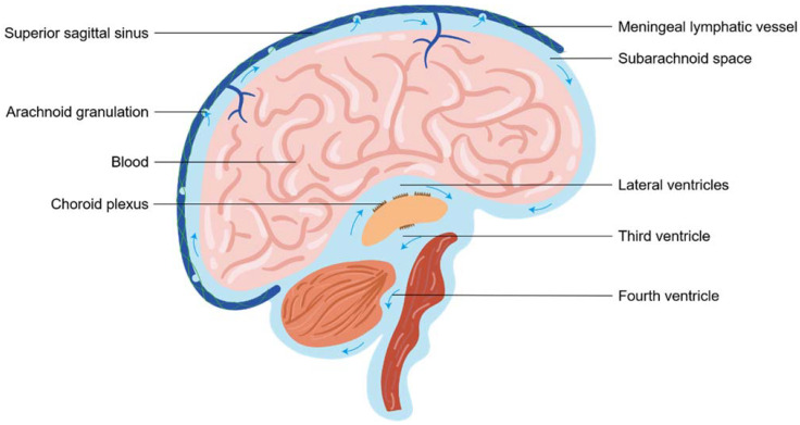
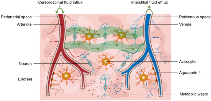
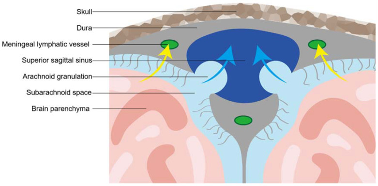
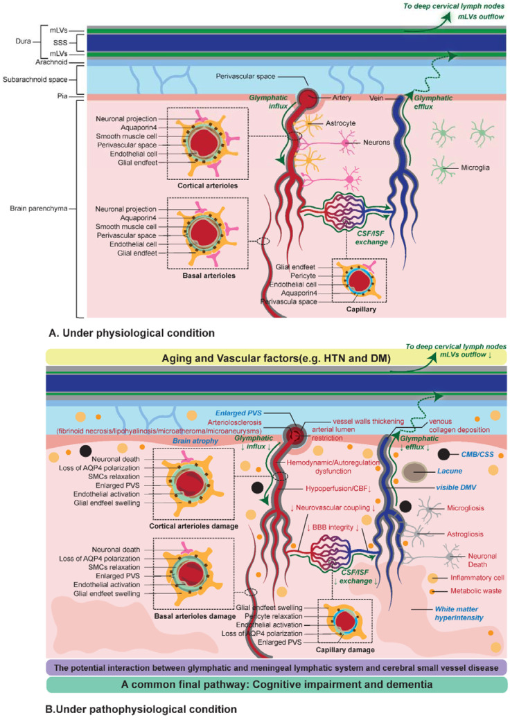
There is a growing prevalence of vascular cognitive impairment (VCI) worldwide, and most research has suggested that cerebral small vessel disease (CSVD) is the main contributor to VCI. Several potential physiopathologic mechanisms have been proven to be involved in the process of CSVD, such as blood-brain barrier damage, small vessels stiffening, venous collagenosis, cerebral blood flow reduction, white matter rarefaction, chronic ischaemia, neuroinflammation, myelin damage, and subsequent neurodegeneration. However, there still is a limited overall understanding of the sequence and the relative importance of these mechanisms. The glymphatic system (GS) and meningeal lymphatic vessels (mLVs) are the analogs of the lymphatic system in the central nervous system (CNS). As such, these systems play critical roles in regulating cerebrospinal fluid (CSF) and interstitial fluid (ISF) transport, waste clearance, and, potentially, neuroinflammation. Accumulating evidence has suggested that the glymphatic and meningeal lymphatic vessels played vital roles in animal models of CSVD and patients with CSVD. Given the complexity of CSVD, it was significant to understand the underlying interaction between glymphatic and meningeal lymphatic transport with CSVD. Here, we provide a novel framework based on new advances in main four aspects, including vascular risk factors, potential mechanisms, clinical subtypes, and cognition, which aims to explain how the glymphatic system and meningeal lymphatic vessels contribute to the progression of CSVD and proposes a comprehensive insight into the novel therapeutic strategy of CSVD.

Keywords: cerebral small vessel disease; cerebrospinal fluid; glymphatic system; meningeal lymphatic vessel.


Conflict of interest statement

The authors declare no conflict of interest.

Figures

Figure 1
Figure 1
A brief overview of cerebrospinal fluid circulation. Traditionally, Cerebrospinal fluid (CSF) produced in the choroid plexus flows from later ventricles, the third ventricle and fourth ventricle to the subarachnoid space (SAS) of the brain. In the SAS, CSF was reabsorbed through the arachnoid granulations into the venous sinuses for efflux. In addition, the perineural sheaths surrounding cranial nerves through the cribriform plate also could drain CSF. Recent advances suggested meningeal lymphatic vessels drained CSF to deep cervical lymph nodes. The contribution and importance of these CSF outflow pathways are debated.
Figure 2
Figure 2
A brief overview of the glymphatic system. The glymphatic system consisted of three main components: cerebrospinal fluid (CSF) influx along periarterial spaces, exchange between CSF and interstitial fluid (ISF) in the brain parenchyma, and ISF efflux along perivenous spaces. Aquaporin4 (AQP4) located in astrocyte endfeet toward perivascular spaces were essential to maintain the normal function of the glymphatic transport, such as metabolic waste clearance. The loss of AQP4 polarization resulted in glymphatic failure in aging.
Figure 3
Figure 3
A brief overview of meningeal lymphatic vessels. Meningeal lymphatic vessels (mLVs) could be described as a lymphatic network in the central nervous system that expresses classic protein markers of lymphatic endothelial cells, such as lymphatic vessel endothelial hyaluronan receptor 1 (LYVE1), vascular endothelial growth factor receptor 3 (VEGFR3), prospero homeobox protein 1 (Prox1), and chemokine (C-C motif) ligand 21 (CCL21). Developmentally, the maturity of mLVS depend on vascular endothelial growth factor C (VEGF-C). Anatomically, mLVs extend from the superior sagittal sinus and the transverse sinuses to into the deep cervical lymph nodes (dCLNs). Functionally, mLVs play a significant role in draining of CSF, metabolic waste, immune cells, or other substances.
Figure 4
Figure 4
The possible hypothesis of potential interaction between CSVD and glymphatic and meningeal lymphatic system. In sporadic CSVD, normal aging may be an initial event in CSVD pathobiology. Under the conditions of normal aging, vascular risk factors (i.e., hypertension and diabetes), and genetic factors cause a series of structural and functional changes in the brain and cerebral small vessels occur. There are four main aspects of pathological and pathophysiological mechanisms involved in CSVD: cerebral small vascular cellular damage, abnormal structure of cerebral small vessels, abnormal regulation of cerebral small vessels, and cerebral homeostasis imbalance. Although we proposed a structured insight of the pathological and pathophysiological mechanisms of CSVD, in fact, these processes are not independent, and there is a continuous dynamic interaction among these mechanisms. The pathophysiological processes of CSVD were related with glymphatic and meningeal lymphatic failure. Neuroimaging features on MRI are direct evidence of CSVD, including cortical superficial siderosis, cerebral microbleeds, cortical microinfarction, lacunas, white matter hyperintensity, brain atrophy, enlarged perivascular space (EPVS), and deep medullary veins. PVSs are important composite parts of the glymphatic system, whereas EPVSs visible on MRI indicate impaired glymphatic and meningeal lymphatic transport. A vicious circle was created between the glymphatic and meningeal lymphatic dysfunction and CSVD that may finally result in cognitive impairment and dementia.

References

    1. Nam K., Kwon H., Lim J., Han M., Nam H., Lee Y. The presence and severity of cerebral small vessel disease increases the frequency of stroke in a cohort of patients with large artery occlusive disease. PLoS ONE. 2017;12:e184944. doi: 10.1371/journal.pone.0184944. - DOI - PMC - PubMed
    1. Gorelick P.B., Scuteri A., Black S.E., Decarli C., Greenberg S.M., Iadecola C., Launer L.J., Laurent S., Lopez O.L., Nyenhuis D., et al. Vascular contributions to cognitive impairment and dementia: A statement for healthcare professionals from the american heart association/american stroke association. Stroke. 2011;42:2672–2713. doi: 10.1161/STR.0b013e3182299496. - DOI - PMC - PubMed
    1. De Leeuw F.E., de Groot J.C., Achten E., Oudkerk M., Ramos L.M., Heijboer R., Hofman A., Jolles J., van Gijn J., Breteler M.M. Prevalence of cerebral white matter lesions in elderly people: A population based magnetic resonance imaging study. The Rotterdam Scan Study. J. Neurol. Neurosurg. Psychiatry. 2001;70:9–14. doi: 10.1136/jnnp.70.1.9. - DOI - PMC - PubMed
    1. Wardlaw J.M., Smith E.E., Biessels G.J., Cordonnier C., Fazekas F., Frayne R., Lindley R.I., O’Brien J.T., Barkhof F., Benavente O.R., et al. Neuroimaging standards for research into small vessel disease and its contribution to ageing and neurodegeneration. Lancet Neurol. 2013;12:822–838. doi: 10.1016/S1474-4422(13)70124-8. - DOI - PMC - PubMed
    1. Shi Y., Wardlaw J.M. Update on cerebral small vessel disease: A dynamic whole-brain disease. BMJ. 2016;1:83–92. doi: 10.1136/svn-2016-000035. - DOI - PMC - PubMed

Publication types