

The Repeating, Modular Architecture of the HtrA Proteases
- PMID: 35740918
- PMCID: PMC9221053
- DOI: 10.3390/biom12060793
The Repeating, Modular Architecture of the HtrA Proteases
Abstract
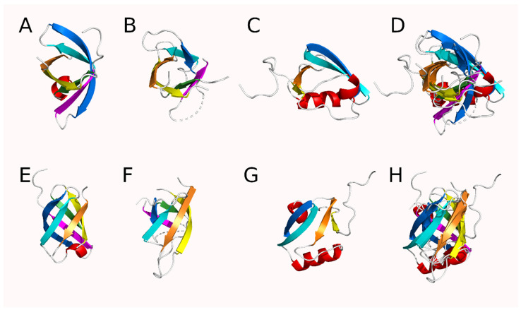
A conserved, 26-residue sequence [AA(X2)[A/G][G/L](X2)GDV[I/L](X2)[V/L]NGE(X1)V(X6)] and corresponding structure repeating module were identified within the HtrA protease family using a non-redundant set (N = 20) of publicly available structures. While the repeats themselves were far from sequence perfect, they had notable conservation to a statistically significant level. Three or more repetitions were identified within each protein despite being statistically expected to randomly occur only once per 1031 residues. This sequence repeat was associated with a six stranded antiparallel β-barrel module, two of which are present in the core of the structures of the PA clan of serine proteases, while a modified version of this module could be identified in the PDZ-like domains. Automated structural alignment methods had difficulties in superimposing these β-barrels, but the use of a target human HtrA2 structure showed that these modules had an average RMSD across the set of structures of less than 2 Å (mean and median). Our findings support Dayhoff's hypothesis that complex proteins arose through duplication of simpler peptide motifs and domains.
Keywords: HtrA protease; PA clan; protein evolution; protein repeat; serine protease.
Conflict of interest statement
The authors declare no conflict of interest.
Figures


Similar articles
-
Peptide selectivity between the PDZ domains of human pregnancy-related serine proteases (HtrA1, HtrA2, HtrA3, and HtrA4) can be reshaped by different halogen probes.J Mol Recognit. 2018 Jun;31(6):e2698. doi: 10.1002/jmr.2698. Epub 2017 Dec 20. J Mol Recognit. 2018. PMID: 29266444
-
Solution structure of HtrA PDZ domain from Streptococcus pneumoniae and its interaction with YYF-COOH containing peptides.J Struct Biol. 2011 Oct;176(1):16-23. doi: 10.1016/j.jsb.2011.06.009. Epub 2011 Jul 2. J Struct Biol. 2011. PMID: 21757011
-
PDZ domains facilitate binding of high temperature requirement protease A (HtrA) and tail-specific protease (Tsp) to heterologous substrates through recognition of the small stable RNA A (ssrA)-encoded peptide.J Biol Chem. 2002 Oct 18;277(42):39443-9. doi: 10.1074/jbc.M202790200. Epub 2002 Aug 12. J Biol Chem. 2002. PMID: 12177052
-
The HtrA family of serine proteases.Mol Microbiol. 1997 Oct;26(2):209-21. doi: 10.1046/j.1365-2958.1997.5601928.x. Mol Microbiol. 1997. PMID: 9383148 Review.
-
HTRA proteases: regulated proteolysis in protein quality control.Nat Rev Mol Cell Biol. 2011 Mar;12(3):152-62. doi: 10.1038/nrm3065. Epub 2011 Feb 16. Nat Rev Mol Cell Biol. 2011. PMID: 21326199 Review.
Cited by
-
Advances in Computational Intelligence-Based Methods of Structure and Function Prediction of Proteins.Biomolecules. 2024 Aug 29;14(9):1083. doi: 10.3390/biom14091083. Biomolecules. 2024. PMID: 39334850 Free PMC article.
-
The NtDEGP5 gene improves drought tolerance in tobacco (Nicotiana tabacum L.) by dampening plastid extracellular Ca2+ and flagellin signaling and thereby reducing ROS production.Plant Mol Biol. 2023 Nov;113(4-5):265-278. doi: 10.1007/s11103-023-01388-8. Epub 2023 Nov 20. Plant Mol Biol. 2023. PMID: 37985581
References
-
- Burley S.K., Berman H.M., Bhikadiya C., Bi C.X., Chen L., Di Costanzo L., Christie C., Dalenberg K., Duarte J.M., Dutta S., et al. RCSB Protein Data Bank: Biological macromolecular structures enabling research and education in fundamental bi-ology, biomedicine, biotechnology and energy. Nucleic Acids Res. 2019;47:D464–D474. doi: 10.1093/nar/gky1004. - DOI - PMC - PubMed
Publication types
MeSH terms
Substances
LinkOut - more resources
Full Text Sources
