. 2022 Jun 9;12(6):807.
doi: 10.3390/biom12060807.

The Applicability of Current Turbidimetric Approaches for Analyzing Fibrin Fibers and Other Filamentous Networks

Affiliations

The Applicability of Current Turbidimetric Approaches for Analyzing Fibrin Fibers and Other Filamentous Networks

Heather A Belcher et al. Biomolecules. .

Abstract

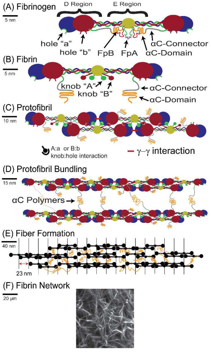
Turbidimetry is an experimental technique often used to study the structure of filamentous networks. To extract structural properties such as filament diameter from turbidimetric data, simplifications to light scattering theory must be employed. In this work, we evaluate the applicability of three commonly utilized turbidimetric analysis approaches, each using slightly different simplifications. We make a specific application towards analyzing fibrin fibers, which form the structural scaffold of blood clots, but the results are generalizable. Numerical simulations were utilized to assess the applicability of each approach across a range of fiber lengths and diameters. Simulation results indicated that all three turbidimetric approaches commonly underestimate fiber diameter, and that the “Carr-Hermans” approach, utilizing wavelengths in the range of 500−800 nm, provided <10% error for the largest number of diameter/length combinations. These theoretical results were confirmed, under select conditions, via the comparison of fiber diameters extracted from experimental turbidimetric data, with diameters obtained using super-resolution microscopy.

Keywords: fibrin; filamentous networks; light scattering; turbidimetry; turbidity.


Conflict of interest statement

The authors declare no conflict of interest.

Figures

Figure 1
Figure 1
Schematic of the polymerization process: (A) Cartoon of the fibrinogen protein: the α chain is shown in green, β chain in red, and γ chain in blue; the disulfide bond-rich center of the molecule, where all six chains are connected, is depicted in yellow. (B) The fibrin molecule: upon thrombin cleavage of FpA and FpB, knob A and knob B are exposed to bind the respective hole a and hole b. (C) A half-staggered protofibril grows longitudinally as the knobs in the central region of one molecule bind to the holes in the distal region of two opposite molecules. (D) Lateral aggregation of protofibrils, likely mediated by interactions of the αC regions, which consist of the αC-connector and αC-domain. (E) Further aggregation of protofibrils into fibrin fibers. (F) A representative image of fibrin fibers in a gel. The image is a maximum intensity projection of a fibrin clot formed from human plasma spiked with 0.1% Alexa-488 labeled fibrinogen, imaged using a Bruker MuVi-SPIM light sheet microscope. Solid black lines on the left of each panel show the scale.
Figure 2
Figure 2
The incoming light enters the solution and is scattered by the rods. The transmitted light is collected by a detector. A ratio of the transmitted light to incoming light can be used to determine the turbidity.
Figure 3
Figure 3
An outline of the methods for determining the percent error in each approach, using constant values of n and dn/dc.
Figure 4
Figure 4
Percent error in diameter (A,B) and in mass–length ratio (C,D) between the values obtained from fitting the Carr–Hermans approach to the full light scattering theory dataset and the values used to create the initial dataset for lengths of 0.5–10 μm and diameters of 10–200 nm. In (A,C), the wavelength dependence of n and dn/dc was accounted for (Equations (20) and (21); in (B,D), constant values for n and dn/dc at 633 nm were used. (c = 0.0001 g/cm3, μ = 4.73 × 10¹² Da/cm, HBS buffer; the purple bars represent imaginary diameter values calculated from the fit and were assigned a value of 100% error for plotting purposes; note that (A,B) have a different viewing angle to (C,D).
Figure 5
Figure 5
Percent error between the diameter obtained from fitting the three approaches to theoretical turbidity values created using full light scattering theory, and the value used to create the initial dataset for lengths of 0.5–10 μm and diameters of 10–200 nm for wavelength ranges of 350–650 nm (AC) and 500–800 nm (DF). (c = 0.0001 g/cm3, μ = 4.73 × 10¹² Da/cm, dn/dc and n corrected for wavelength dependence for fibers in HBS buffer; the purple bars represent imaginary diameter values calculated from the fit, and were assigned a value of 100% error for plotting purposes).
Figure 6
Figure 6
Percent error between the mass–length ratio obtained from fitting the approaches to theoretical turbidity values created using full light scattering theory and the value used to create the initial dataset for lengths of 0.5–10 μm and diameters of 10–200 nm for wavelength ranges of 350–650 nm (A,B) and 500–800 nm (C,D). (c = 0.0001 g/cm3, μ = 4.73 × 10¹² Da/cm, dn/dc and n corrected for wavelength dependence for fibers in HBS buffer).
Figure 7
Figure 7
Stochastic optical reconstruction microscopy (STORM) image of a clot formed from 0.5 mg/mL fibrinogen and 0.1 NIH-U/mL thrombin (in HBS buffer + 5 mM calcium chloride), with AlexaFluor-647 labeled fibrinogen added at a concentration of 1/65 that of the wild-type fibrinogen to allow for fluorescence imaging, obtained on a Nikon Ti2-E inverted microscope using a 100× oil objective.

Similar articles

Cited by

References

    1. Zimm B.H., Dandliker W.B. Theory of Light Scattering and Refractive Index of Solutions of Large Colloidal Particles. J. Phys. Chem. 1954;58:644–648. doi: 10.1021/j150518a012. - DOI
    1. Kurniawan N.A., Grimbergen J., Koopman J., Koenderink G.H. Factor XIII stiffens fibrin clots by causing fiber compaction. J. Thromb. Haemost. 2014;12:1687–1696. doi: 10.1111/jth.12705. - DOI - PubMed
    1. Morozova S., Muthukumar M. Electrostatic effects in collagen fibril formation. J. Chem. Phys. 2018;149:163333. doi: 10.1063/1.5036526. - DOI - PubMed
    1. Rangel-Argote M., Claudio-Rizo J.A., Castellano L.E., Vega-González A., Mata-Mata J.L., Mendoza-Novelo B. ECM-oligourethene-silica hydrogels as a local drug release system of dexamethasone for stimulating macrophages. RSC Adv. 2017;17:10443–10453. doi: 10.1039/C6RA25989H. - DOI
    1. Zhu J., Kaufman L.J. Collagen I Self-Assembly: Revealing the Developing Structures that Generate Turbidity. Biophys. J. 2014;106:1822–1831. doi: 10.1016/j.bpj.2014.03.011. - DOI - PMC - PubMed

Publication types