. 2022 Jun 12;12(6):820.
doi: 10.3390/biom12060820.

Occurrence of Lymphangiogenesis in Peripheral Nerve Autografts Contrasts Schwann Cell-Induced Apoptosis of Lymphatic Endothelial Cells In Vitro

Affiliations

Occurrence of Lymphangiogenesis in Peripheral Nerve Autografts Contrasts Schwann Cell-Induced Apoptosis of Lymphatic Endothelial Cells In Vitro

Carina Hromada et al. Biomolecules. .

Abstract

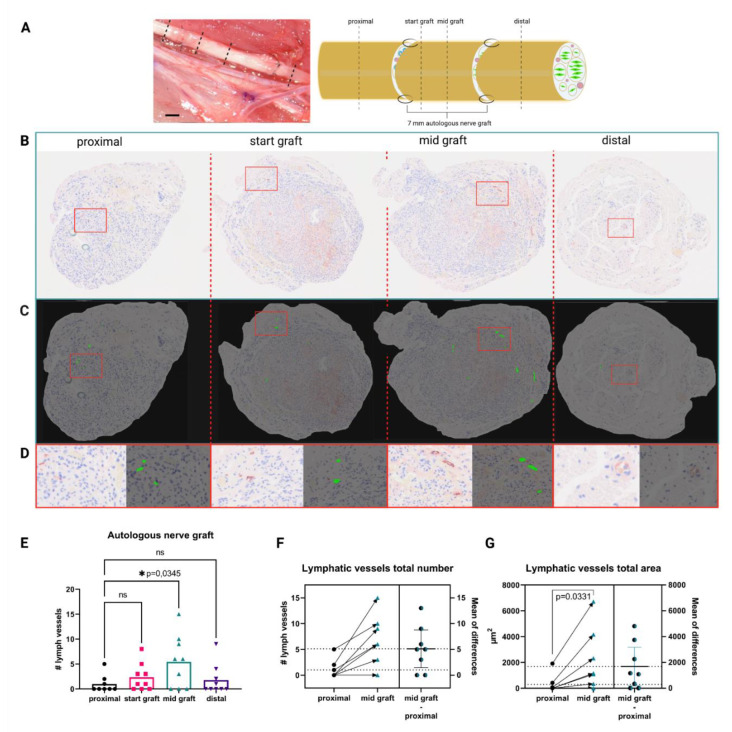
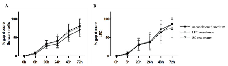
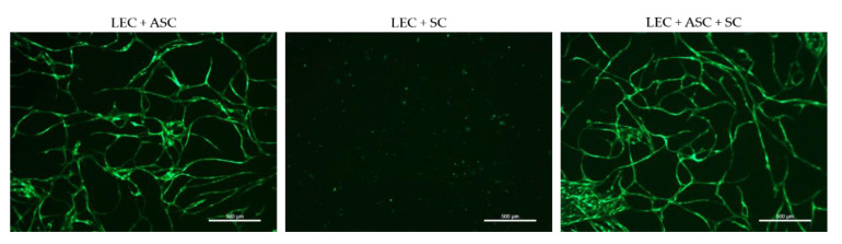
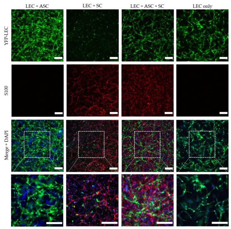
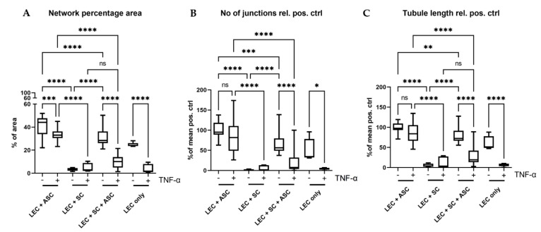
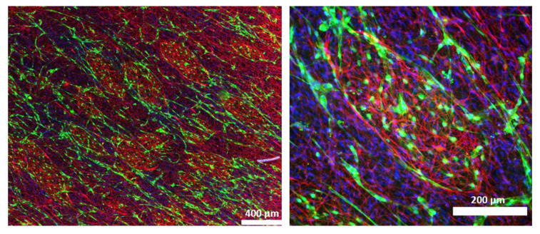
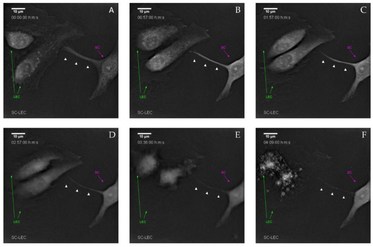
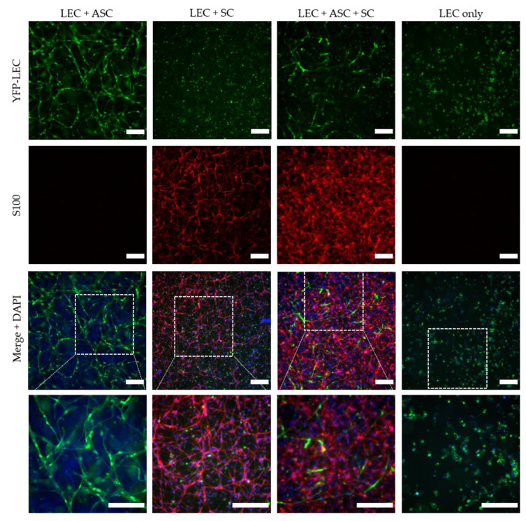
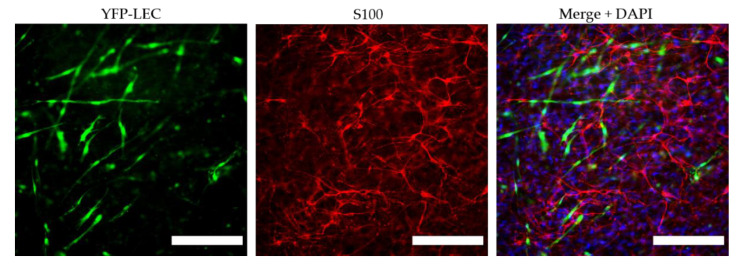
Peripheral nerve injuries pose a major clinical concern world-wide, and functional recovery after segmental peripheral nerve injury is often unsatisfactory, even in cases of autografting. Although it is well established that angiogenesis plays a pivotal role during nerve regeneration, the influence of lymphangiogenesis is strongly under-investigated. In this study, we analyzed the presence of lymphatic vasculature in healthy and regenerated murine peripheral nerves, revealing that nerve autografts contained increased numbers of lymphatic vessels after segmental damage. This led us to elucidate the interaction between lymphatic endothelial cells (LECs) and Schwann cells (SCs) in vitro. We show that SC and LEC secretomes did not influence the respective other cell types' migration and proliferation in 2D scratch assay experiments. Furthermore, we successfully created lymphatic microvascular structures in SC-embedded 3D fibrin hydrogels, in the presence of supporting cells; whereas SCs seemed to exert anti-lymphangiogenic effects when cultured with LECs alone. Here, we describe, for the first time, increased lymphangiogenesis after peripheral nerve injury and repair. Furthermore, our findings indicate a potential lymph-repellent property of SCs, thereby providing a possible explanation for the lack of lymphatic vessels in the healthy endoneurium. Our results highlight the importance of elucidating the molecular mechanisms of SC-LEC interaction.

Keywords: Schwann cells; lymphangiogenesis; lymphatic endothelial cells; peripheral nerve regeneration.


Conflict of interest statement

The authors declare no conflict of interest. The funders had no role in the design of the study; in the collection, analyses, or interpretation of data; in the writing of the manuscript, or in the decision to publish the results.

Figures

Figure 1
Figure 1
Autologous nerve grafting results in increased intraneural lymphangiogenesis. (A) in vivo appearance of the median nerve after autologous nerve grafting and schematic of the repaired median nerve, with histological sections and their respective localization indicated as proximal, start graft, mid graft, and distal. Dotted lines indicate histological cutting planes. Scale bar = 1 mm (B) Representative histological cross sections of the rat median nerve, 3 months after autologous nerve grafting stained for podoplanin. From left to right: proximal to the autologous nerve graft, start of the graft, mid-section of the graft, and distal to the graft; red rectangles indicate enlarged regions of interest depicted in (D). (C) Readout of the automated identification using KML Vision IKOSA deep learning algorithm, lymphatic vessels are highlighted in green, red rectangles indicate enlarged regions of interest depicted in (D). (D) Side-by-side comparison of respective regions of interest within histological pictures with automatically identified lymphatic vessels. (E) Quantification of podoplanin+ lymphatic vessels in the histological sections shows a significantly higher number of lymphatic vessels at mid graft level, when compared to the proximal nerve section. (F) Comparison of lymphatic vessel number proximal to mid graft in individual nerves shows increases in lymphatic vessel number in almost all samples. The average increase in vessel number from proximal to mid graft was 5; lower dotted line indicates the mean lymphatic vessel number in the proximal segment; whereas, the upper dotted line indicates the mean lymphatic vessel number in the mid graft segment. Arrows indicate changes in nerve segments of the same samples (n = 8). (G) Similar to (F), the comparison of the total vessel area also revealed a significant increase from the proximal to the mid graft segment of individual nerves, with an average increase of 1800 µm2, dotted lines represent average proximal and mid graft lymphatic vessel area (n = 8). (ns—not significant, * p < 0.05 was considered significant, asterisk indicates significance, matched one-way ANOVA with Dunnet’s test for multiple comparison was used to compare lymphatic vessel numbers in different nerve segments to the proximal nerve segment, a paired t-test was performed to compare proximal and mid graft lymphatic vessel total area).
Figure 2
Figure 2
Schwann cell migration was not altered by lymphatic endothelial cell secretome in 2D scratch assay experiments. SCs and LECs were grown until confluency and their conditioned media were collected 24 h before the experiment. Scratches were created on cell monolayers and cell migration in either unconditioned medium or conditioned medium from LECs or SCs was monitored over a total of 72 h. (A,B) Quantification of gap closure showed that the secretome of LECs and SCs did not affect the migration of either SCs or LECs. Data are presented as mean ± sd. LEC secretome and SC secretome: n = 15 of 3 independent experiments; unconditioned medium: n = 12 of 2 independent experiments.
Figure 3
Figure 3
2D culture of YFP-LECs (green) with either ASCs or SCs alone, or with both ASCs and SCs. Cells were seeded in equal densities. Elongated lymphatic network-like structures formed when LECs were co-cultured with ASCs as well as with both ASCs and SCs. In contrast, LECs remained as single cells but also seemed to decrease in number when cultured with SCs alone. Green—YFP-LECs. Scale bars = 500 µm.
Figure 4
Figure 4
Lymphatic network formation is decreased in the presence of Schwann cells in 3D fibrin hydrogels. LECs formed elongated and interconnected networks when cultivated with ASCs as a supporting cell type (LEC + ASC). In a tri-culture of LECs, ASCs, and SCs, lymphatic vessel formation was less dense and interconnected (LEC + ASC + SC), whereas in the monoculture of LECs, only primitive tube-like structures were formed. In contrast, co-culture of only LECs and SCs inhibited the formation of lymphatic tubes. Green—YFP-LECs, red—SCs stained against S100, blue—cell nuclei. Scale bars = 200 µm.
Figure 5
Figure 5
Quantification of the formed lymphatic networks in 3D fibrin clots. (A) Evaluation of the average area covered by lymphatic networks in percentage of total area. (B) Evaluation of the average number of junctions relative to the mean of the positive control (LEC + ASC). (C) Quantification of the average lymphatic tubule length relative to the mean of the positive control (LEC + ASC). In all parameters, the triple culture of LECs, SCs, and ASCs showed significantly lower levels than the positive control (LEC + ASC) (AC). Co-culture of LECs with SCs inhibited network formation, whereas addition of ASCs reversed this effect. Furthermore, the effect of pro-inflammatory TNF-α on the formation of lymphatic networks was also evaluated. Here, we observed a larger negative effect of TNF-α on the triple-culture, when compared to the positive control in all parameters. LEC + ASC, LEC + SC and LEC + ASC + SC: n = 8 each of 4 independent experiments, LEC only: n = 4 of 2 independent experiments. * p < 0.05, ** p < 0.01, *** p < 0.001, **** p < 0.0001, ns not significant.
Figure 6
Figure 6
Schwann cells (red) assemble into islands, which are surrounded by lymphatic vessels (green) in co-cultures of LECs, ASCs, and SCs. Green—YFP-LECs, red—SCs stained against S100, blue—cell nuclei.
Figure 7
Figure 7
Live cell imaging demonstrates that cell–cell contact induced lymphatic endothelial cell death by Schwann cells. (A) Initial cell–cell contact between LECs (green arrow) and SCs (magenta arrow) via filopodia-like protrusions (white arrow heads). (B) After initial contact, morphological changes in both LECs and the SC-filopodia-like protrusions become visible. (C,D) Retraction of both the filopodia-like protrusion and LEC endoplasm. (E) Stark retraction and compartmentalization of LECs. (F) Formation of LEC-derived apoptotic bodies. Time-lapse shown in each image is the observation time starting 2 h post-seeding.
Figure 8
Figure 8
Schwann cells hinder lymphatic vascular network formation in the presence of the pro-inflammatory cytokine TNF-α. LEC + ASC formed elongated and interconnected networks, even when stimulated with TNF-α; whereas the formation of lymphatic vessel-like structures in the LEC + ASC + SC cultures in the presence of TNF-α was hampered. The co-culture of only LECs and SCs did not show the formation of lymphatic tubes. Green—YFP-LECs, red—SCs stained against S100, blue—cell nuclei. Scale bars = 200 µm.
Figure 9
Figure 9
TNF-α induces phenotypical changes in Schwann cells. Stimulation with TNF-α resulted in a morphological change of SCs from a bipolar to a multipolar phenotype, with highly branched protrusions. Green—YFP-LECs, red—SCs stained against S100, blue—cell nuclei. Scale bars = 200 µm.

Similar articles

Cited by

References

    1. Foster C.H., Karsy M., Jensen M.R., Guan J., Eli I., A Mahan M. Trends and Cost-Analysis of Lower Extremity Nerve Injury Using the National Inpatient Sample. Neurosurgery. 2018;85:250–256. doi: 10.1093/neuros/nyy265. - DOI - PubMed
    1. Karsy M., Watkins R., Jensen M.R., Guan J., Brock A.A., Mahan M.A. Trends and Cost Analysis of Upper Extremity Nerve Injury Using the National (Nationwide) Inpatient Sample. World Neurosurg. 2018;123:e488–e500. doi: 10.1016/j.wneu.2018.11.192. - DOI - PubMed
    1. López-Cebral R., Silva-Correia J.S., Reis R.L., Silva T.H., Oliveira J.M. Peripheral Nerve Injury: Current Challenges, Conventional Treatment Approaches, and New Trends in Biomaterials-Based Regenerative Strategies. ACS Biomater. Sci. Eng. 2017;3:3098–3122. doi: 10.1021/acsbiomaterials.7b00655. - DOI - PubMed
    1. Grinsell D., Keating C.P. Peripheral Nerve Reconstruction after Injury: A Review of Clinical and Experimental Therapies. BioMed Res. Int. 2014;2014:698256. doi: 10.1155/2014/698256. - DOI - PMC - PubMed
    1. Heinzel J.C., Dadun L.F., Prahm C., Winter N., Bressler M., Lauer H., Ritter J., Daigeler A., Kolbenschlag J. Beyond the Knife—Reviewing the Interplay of Psychosocial Factors and Peripheral Nerve Lesions. J. Pers. Med. 2021;11:1200. doi: 10.3390/jpm11111200. - DOI - PMC - PubMed

Publication types