. 2022 Jun 19;12(6):849.
doi: 10.3390/biom12060849.

Excess Heme Promotes the Migration and Infiltration of Macrophages in Endometrial Hyperplasia Complicated with Abnormal Uterine Bleeding

Affiliations

Excess Heme Promotes the Migration and Infiltration of Macrophages in Endometrial Hyperplasia Complicated with Abnormal Uterine Bleeding

Lu-Yu Ruan et al. Biomolecules. .

Abstract

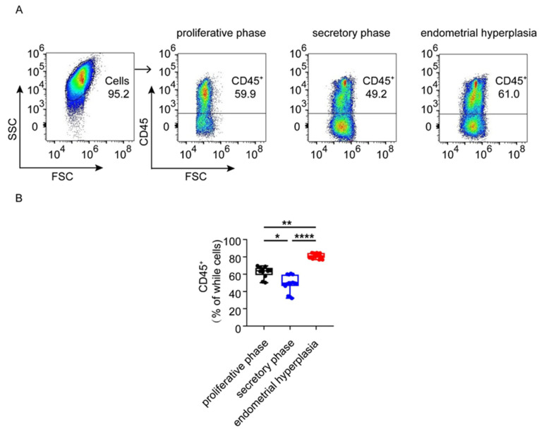
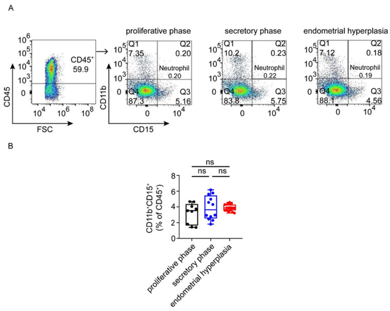
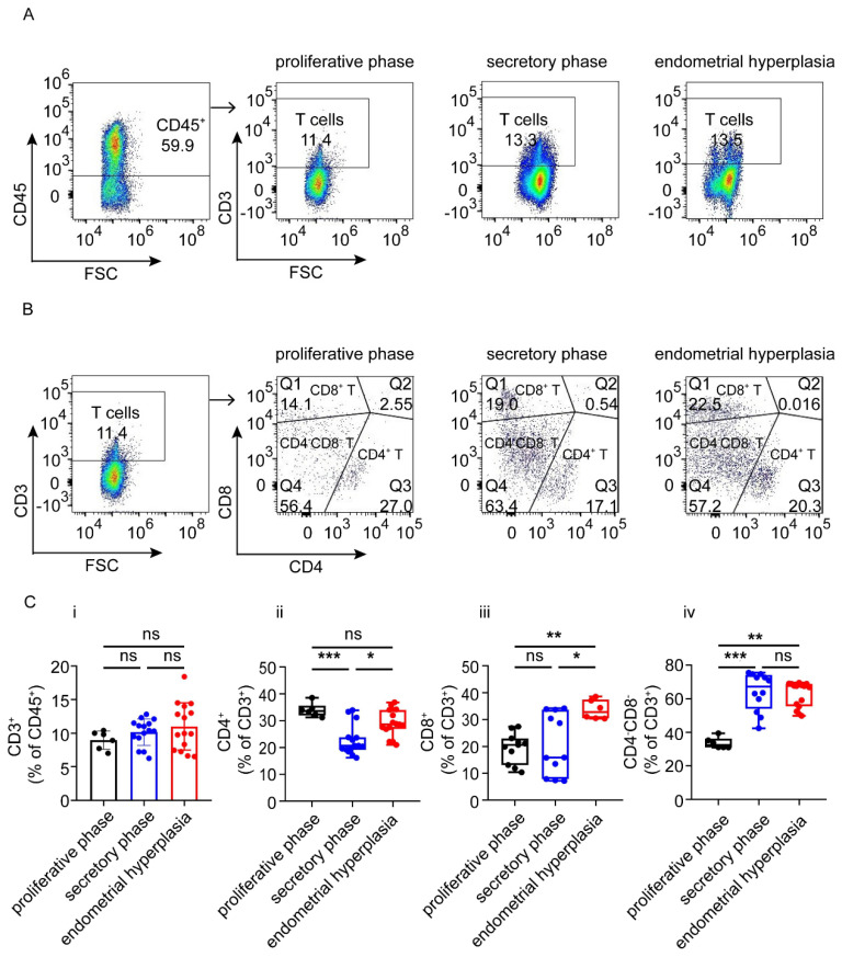
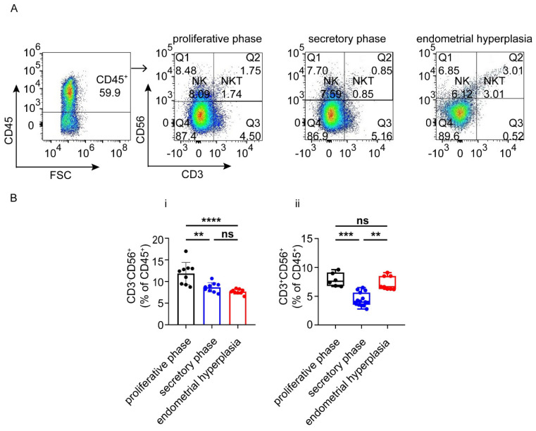
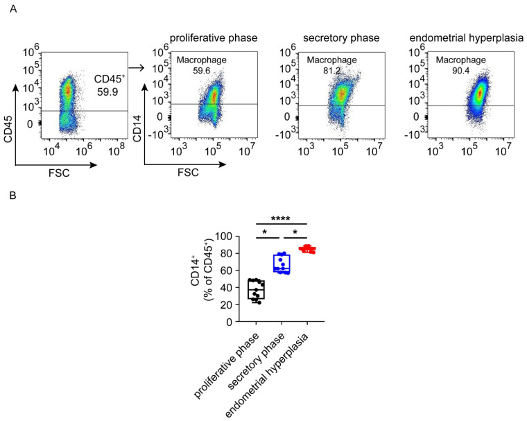
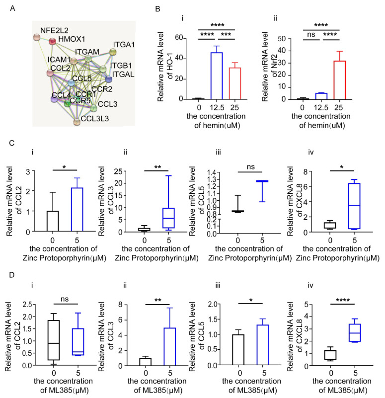
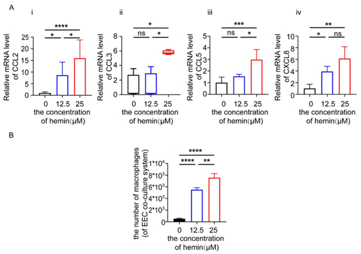
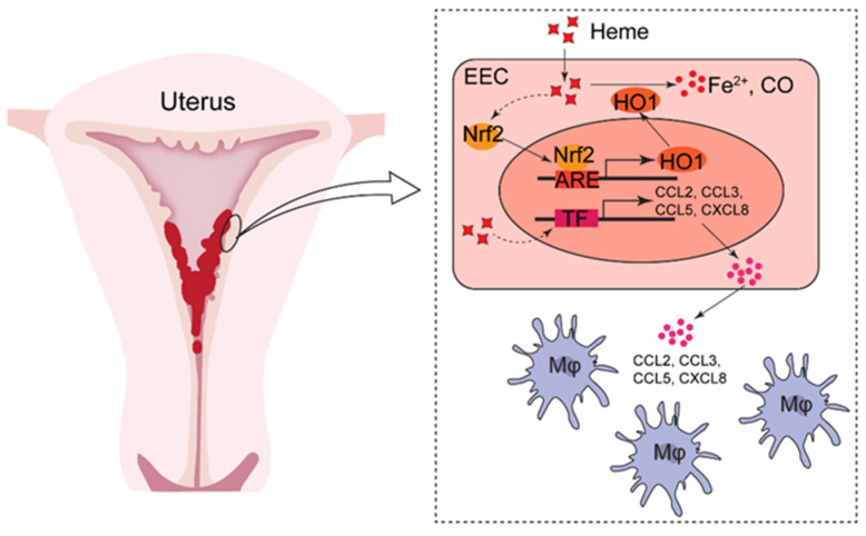
In patients, endometrial hyperplasia (EH) is often accompanied by abnormal uterine bleeding (AUB), which is prone to release large amounts of heme. However, the role of excess heme in the migration and infiltration of immune cells in EH complicated by AUB remains unknown. In this study, 45 patients with AUB were divided into three groups: a proliferative phase group (n = 15), a secretory phase group (n = 15) and EH (n = 15). We observed that immune cell subpopulations were significantly different among the three groups, as demonstrated by flow cytometry analysis. Of note, there was a higher infiltration of total immune cells and macrophages in the endometrium of patients with EH. Heme up-regulated the expression of heme oxygenase-1 (HO-1) and nuclear factor erythroid-2-related factor 2 (Nrf2) in endometrial epithelial cells (EECs) in vitro, as well as chemokine (e.g., CCL2, CCL3, CCL5, CXCL8) levels. Additionally, stimulation with heme led to the increased recruitment of THP-1 cells in an indirect EEC-THP-1 co-culture unit. These data suggest that sustained and excessive heme in patients with AUB may recruit macrophages by increasing the levels of several chemokines, contributing to the accumulation and infiltration of macrophages in the endometrium of EH patients, and the key molecules of heme metabolism, HO-1 and Nrf2, are also involved in this regulatory process.

Keywords: HO-1; abnormal uterine bleeding; endometrial hyperplasia; heme; immune cells; macrophages.


Conflict of interest statement

The authors declare no financial or commercial conflict of interest.

Figures

Figure 1
Figure 1
The numbers of local immunocytes were different in proliferative phase, secretory phase and endometrial hyperplasia endometrium of AUB patients. (A) Using flow cytometry gating strategy to distinguish CD45+ cells in proliferative phase, secretory phase and endometrial hyperplasia endometrium of AUB patients. (B) The proportion of CD45+ cells in whole cells. Data are presented as the median and the interquartile range. Statistical significance (Kruskal–Wallis test with Dunn’s multiple-comparison test): * p < 0.05, ** p < 0.01, **** p < 0.0001.
Figure 2
Figure 2
The proportion of neutrophils in proliferative phase, secretory phase and endometrial hyperplasia endometrium of AUB patients. (A,B) Gating strategy was used to distinguish neutrophils. CD45+ gate was first used to gate cells, followed by CD11b and CD15 gates. Graph shows the proportion of total CD45+CD11b+CD15+ neutrophils in proliferative phase, secretory phase and endometrial hyperplasia groups. Data are presented as the median and the interquartile range. Statistical significance (Kruskal–Wallis test with Dunn’s multiple-comparison test): ns, no significant difference.
Figure 3
Figure 3
Different subtypes of T cells in proliferative phase, secretory phase and endometrial hyperplasia endometrium of AUB patients. (A) CD45+ gate, followed by CD3+ gate, was used to distinguish T cells. (B) Gating strategy was used to distinguish CD4+ T cells, CD8+ T cells and CD4CD8 T cells. (C) Graph shows the proportion of total T cells, CD4+ T cells, CD8+ T cells and CD4CD8 T cells in proliferative phase, secretory phase endometrium and endometrial hyperplasia endometrium of AUB patients. Data are presented as the mean ± standard error of the mean (C.i), or the median and the interquartile range (C.iiC.iv). Statistical significance (one-way ANOVA with a Bonferroni multiple-comparisons test (C.i) or Kruskal–Wallis test with Dunn’s multiple-comparisons test (C.iiC.iv)):, * p < 0.05, ** p < 0.01, *** p < 0.001, ns, no significant difference.
Figure 4
Figure 4
NK and NKT cells in proliferative phase, secretory phase and endometrial hyperplasia endometrium of AUB patients. (A) CD45+ gate, followed by CD3+/−CD56+ gate, was used to distinguish NK cells and NKT cells. (B) Graph shows the proportions of general NK cells and NKT cells in three groups. Data are presented as the mean ± standard error of the mean (B.i), or the median and the interquartile range (B.ii). Statistical significance (one-way ANOVA with a Bonferroni multiple-comparisons test (B.i) or Kruskal–Wallis test with Dunn’s multiple-comparisons test (B.ii)): ** p < 0.01, *** p < 0.001, **** p < 0.0001, ns, no significant difference.
Figure 5
Figure 5
The proportion of macrophages in proliferative phase, secretory phase and endometrial hyperplasia endometrium of AUB patients. (A) CD45+ gate, followed by CD14+ gate, was used to distinguish monocytes. (B) Graph shows the proportions of general monocytes in three groups. Data are presented as the median and the interquartile range. Statistical significance (Kruskal–Wallis test with Dunn’s multiple-comparison test): * p < 0.05, **** p < 0.0001.
Figure 6
Figure 6
Blocking HO-1 and Nrf2 leads to an increase in chemokines by EECs. (A) The PPI network of chemokines, adhesion molecules and heme metabolism-related factors. (B) Relative mRNA expression of HO-1 and Nrf2 in EECs treated with Hemin at a concentration of 0 μM, 12.5 μM and 25 μM for 48 h. (C) Relative mRNA expression of CCL2, CCL3, CCL5, CXCL8 in EECs treated with HO-1 inhibitor (Zinc Protoporphyrin) at the concentration of 5 μM for 24 h. (D) Relative mRNA expression of CCL2, CCL3, CCL5, CXCL8 in EECs treated with Nrf2 inhibitor (ML385) at the concentration of 5 μM for 24 h. Data are presented as the mean ± standard error of the mean (B.i,B.ii,C.i,D.ii,D.iii), or the median and the interquartile range (C.iiC.iv,D.i,D.iv). Statistical significance (one-way ANOVA with a Bonferroni multiple-comparisons test for three groups (B.i,B.ii), and t test (C.i,D.ii,D.iii) or Mann–Whitney U test (C.iiC.iv,D.i,D.iv) for two groups): * p < 0.05, ** p < 0.01, *** p < 0.001, **** p < 0.0001, ns: no significance.
Figure 7
Figure 7
Heme increases the expression of chemokines in EECs and recruits macrophages. (A) Relative mRNA expression of CCL2, CCL3, CCL5, CXCL8 in EECs treated with Hemin at the concentration of 12.5 μM and 25 μM for 48 h. (B) We illustrate the recruitment of macrophages co-cultured with EECs, which were untreated or treated with Hemin at the concentration of 12.5 μM or 25 μM for 48 h; we counted the number of recruited macrophages. Data are presented as the mean ± standard error of the mean (A.i,A.iii,A.iv,B), or the median and the interquartile range (A.ii). Statistical significance (one-way ANOVA with a Bonferroni multiple-comparisons test (A.i,A.iii,A.iv,B) or Kruskal–Wallis test with Dunn’s multiple-comparisons test (A.ii)): * p < 0.05, ** p < 0.01, *** p < 0.001, **** p < 0.0001, ns: no significance.
Figure 8
Figure 8
A schematic chart of excess heme in the migration and infiltration of macrophages from AUB patients with EH. In endometria of patients with EH complicated by abnormal uterine bleeding, there should be massive heme release. A certain level of heme activates the Nrf2/HO-1 axis, and further promotes heme metabolism. However, excess heme stimulates EECs to produce more chemokines (e.g., CCL2, CCL3, CCL5), possibly leading to the migration and infiltration of macrophages in the endometrium and accelerating the development of EH.

Similar articles

Cited by

References

    1. Ottnad E., Parthasarathy S., Sambrano G.R., Ramprasad M.P., Quehenberger O., Kondratenko N., Green S., Steinberg D. A macrophage receptor for oxidized low density lipoprotein distinct from the receptor for acetyl low density lipoprotein: Partial purification and role in recognition of oxidatively damaged cells. Proc. Natl. Acad. Sci. USA. 1995;92:1391–1395. doi: 10.1073/pnas.92.5.1391. - DOI - PMC - PubMed
    1. Ozdegirmenci O., Kayikcioglu F., Bozkurt U., Akgul M.A., Haberal A. Comparison of the efficacy of three progestins in the treatment of simple endometrial hyperplasia without atypia. Gynecol. Obstet. Invest. 2011;72:10–14. doi: 10.1159/000321390. - DOI - PubMed
    1. Sanderson P.A., Critchley H.O., Williams A.R., Arends M.J., Saunders P.T. New concepts for an old problem: The diagnosis of endometrial hyperplasia. Hum. Reprod. Update. 2017;23:232–254. doi: 10.1093/humupd/dmw042. - DOI - PMC - PubMed
    1. Lai Z.Z., Ruan L.Y., Wang Y., Yang H.L., Shi J.W., Wu J.N., Qiu X.M., Ha S.Y., Shen H.H., Yang S.L., et al. Changes in subsets of immunocytes in endometrial hyperplasia. Am. J. Reprod. Immunol. 2020;84:e13295. doi: 10.1111/aji.13295. - DOI - PubMed
    1. Acmaz G., Aksoy H., Unal D., Ozyurt S., Cingillioglu B., Aksoy U., Muderris I. Are neutrophil/lymphocyte and platelet/lymphocyte ratios associated with endometrial precancerous and cancerous lesions in patients with abnormal uterine bleeding? Asian Pac. J. Cancer Prev. 2014;15:1689–1692. doi: 10.7314/APJCP.2014.15.4.1689. - DOI - PubMed

Publication types