

Mesenchymal Stem Cells in Embryo-Maternal Communication under Healthy Conditions or Viral Infections: Lessons from a Bovine Model
- PMID: 35740987
- PMCID: PMC9221285
- DOI: 10.3390/cells11121858
Mesenchymal Stem Cells in Embryo-Maternal Communication under Healthy Conditions or Viral Infections: Lessons from a Bovine Model
Abstract
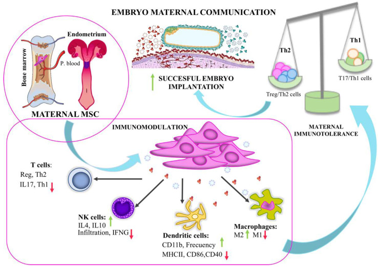
Bovine mesenchymal stem cells are a relevant cell population found in the maternal reproductive tract that exhibits the immunomodulation capacity required to prevent embryo rejection. The phenotypic plasticity showed by both endometrial mesenchymal stem cells (eMSC) and embryonic trophoblast through mesenchymal to epithelial transition and epithelial to mesenchymal transition, respectively, is essential for embryo implantation. Embryonic trophoblast maintains active crosstalk via EVs and soluble proteins with eMSC and peripheral blood MSC (pbMSC) to ensure the retention of eMSC in case of pregnancy and induce the chemotaxis of pbMSC, critical for successful implantation. Early pregnancy-related proteins and angiogenic markers are detected as cargo in EVs and the soluble fraction of the embryonic trophectoderm secretome. The pattern of protein secretion in trophectoderm-EVs changes depending on their epithelial or mesenchymal phenotype and due to the uptake of MSC EVs. However, the changes in this EV-mediated communication between maternal and embryonic MSC populations infected by viruses that cause abortions in cattle are poorly understood. They are critical in the investigation of reproductive viral pathologies.
Keywords: early pregnancy; embryo; extracellular vesicles; mesenchymal stromal cells; trophectoderm.
Conflict of interest statement
The authors declare no conflict of interest.
Figures

References
-
- Lopera-Vasquez R., Hamdi M., Fernandez-Fuertes B., Maillo V., Beltrán-Breña P., Calle A., Redruello A., López-Martín S., Gutierrez-Adan A., Yáñez-Mó M., et al. Extracellular Vesicles from BOEC in In Vitro Embryo Development and Quality. PLoS ONE. 2016;11:e0148083. doi: 10.1371/journal.pone.0148083. - DOI - PMC - PubMed
-
- Mazzarella R., Bastos N.M., Bridi A., del Collado M., Andrade G.M., Pinzon J., Prado C.M., Silva L.A., Meirelles F.V., Pugliesi G., et al. Changes in Oviductal Cells and Small Extracellular Vesicles miRNAs in Pregnant Cows. Front. Vet. Sci. 2021;8:639752. doi: 10.3389/fvets.2021.639752. - DOI - PMC - PubMed
Publication types
MeSH terms
LinkOut - more resources
Full Text Sources
Medical
