. 2022 Jun 7;11(12):1863.
doi: 10.3390/cells11121863.

BECLIN-1-Mediated Autophagy Suppresses Silica Nanoparticle-Induced Testicular Toxicity via the Inhibition of Caspase 8-Mediated Cell Apoptosis in Leydig Cells

Affiliations

BECLIN-1-Mediated Autophagy Suppresses Silica Nanoparticle-Induced Testicular Toxicity via the Inhibition of Caspase 8-Mediated Cell Apoptosis in Leydig Cells

Qianru Zhang et al. Cells. .

Abstract

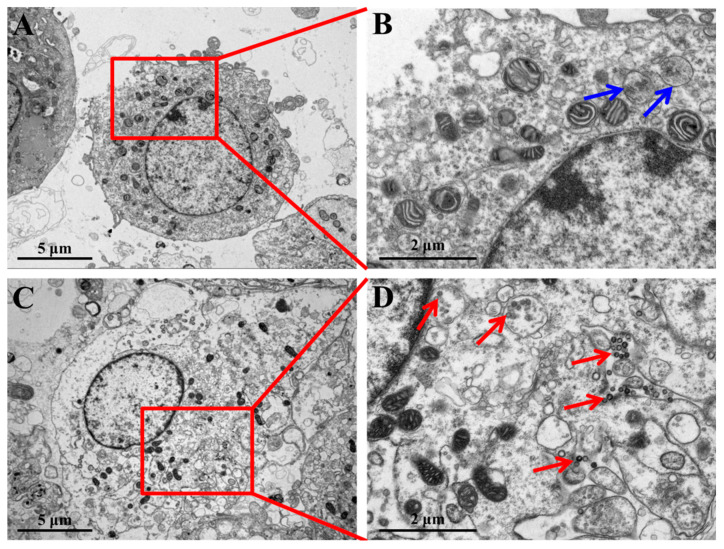
Accumulation of silica nanoparticles (SNPs) in the testes leads to male reproductive toxicity. However, little is known about the effect and mechanistic insights of SNP-induced autophagy on apoptosis in Leydig cells. In this study, we aimed to verify the role of SNP-induced autophagy in apoptosis and explore the possible underlying mechanism in mouse primary Leydig cells (PLCs). H&E staining showed that SNPs changed the histological structures of the testes, including a reduction in the Leydig cell populations in vivo. CCK-8 assay showed that SNPs decreased cell viability, and flow cytometry showed that SNPs increased cell apoptosis, both in a dose-dependent manner in vitro. Additionally, Western blotting further found that SNPs activated autophagy by an increase in BECLIN-1, ATG16L, and LC3-II levels and promoted the intrinsic pathway of apoptosis by an increase in the BAX/BCL-2 ratio, cleaved the caspase 8 and caspase 3 levels. Furthermore, autophagy decreased SNP-induced apoptosis via regulation of the caspase 8 level combined with rapamycin, 3-methyladenine, and chloroquine. BECLIN-1 depletion increased the caspase 8 level, leading to an increase in SNP-induced cell apoptosis. Collectively, this evidence demonstrates that SNPs activated BECLIN-1-mediated autophagy, which prevented SNP-induced testicular toxicity via the inhibition of caspase 8-mediated cell apoptosis in Leydig cells.

Keywords: BECLIN-1; Leydig cells; apoptosis; autophagy; silica nanoparticles; testicular toxicity.


Conflict of interest statement

The authors declare no conflict of interest.

Figures

Figure 1
Figure 1
The morphology and size of SNPs. (A) Representative TEM image of spherical SNPs. Scale bar, 0.5 μm. (B) Distribution diagram of SNPs in (A).
Figure 2
Figure 2
Effects of SNPs on the histological structures of the testes in vivo. (A) The image of the testes in the control group by H&E staining. (B) The selected red area in (A). (C) The selected red area in B. (D) The selected red area in (C). (E) The image of the testes in the SNP-treated group by H&E staining. (F) The selected red area in (E). (G) The selected red area in (F). (H) The selected red area in G. The red arrow represents the spermatogenic cells of the seminiferous tubules; the blue asterisk represents Leydig cells. Scale bars, 200 μm (A,E), 100 μm (B,F), 50 μm (C,G), and 20 μm (D,H).
Figure 3
Figure 3
TEM images of cellular uptake of SNPs. (A) PLCs treated with 400 µg/mL SNPs for 12 h. (B) The selected red square area in (A). (C) Control PLCs. (D) The selected red square area in (C). The red arrows indicate the vesicles where SNPs were enclosed. The blue arrows indicate the vesicles where SNPs were not enclosed. Scale bar, 5 µm (A,C) and 2 µm (B,D).
Figure 4
Figure 4
Effect on the viability and apoptosis after SNP treatment. (A) Cell viability was determined by CCK-8. The PLCs were treated with control, 100, 200, 400, 600, 800, 1000, and 1200 μg/mL SNPs for 24 h. (B) The apoptotic rate was detected by flow cytometry. The PLCs were treated with control, 200, 400, and 800 μg/mL SNPs for 24 h. (C) Quantification and statistical analysis of the apoptotic rate. Note: * p < 0.05, ** p < 0.01, and *** p < 0.001 compared to the control.
Figure 5
Figure 5
Effect of SNPs on the activation of autophagy and the apoptotic pathway. (A) Autophagy was determined by MDC staining. Scale bar, 50 μm (AaAd) and 10 μm (AeAh). (B) Autophagy-related proteins were detected by Western blotting. (C) Bar graphs of the analyses of band intensity in (B) as the relative ratio of the proteins related to β-actin. (D) Apoptosis-related proteins were detected by Western blotting. The PLCs were treated with control, 200, 400, and 800 μg/mL SNPs for 12 h. (E) Bar graphs of the statistical analyses of band intensity in (D) as the relative ratio of the proteins related to β-actin. Note: * p < 0.05, ** p < 0.01, and *** p < 0.001 compared to the control.
Figure 5
Figure 5
Effect of SNPs on the activation of autophagy and the apoptotic pathway. (A) Autophagy was determined by MDC staining. Scale bar, 50 μm (AaAd) and 10 μm (AeAh). (B) Autophagy-related proteins were detected by Western blotting. (C) Bar graphs of the analyses of band intensity in (B) as the relative ratio of the proteins related to β-actin. (D) Apoptosis-related proteins were detected by Western blotting. The PLCs were treated with control, 200, 400, and 800 μg/mL SNPs for 12 h. (E) Bar graphs of the statistical analyses of band intensity in (D) as the relative ratio of the proteins related to β-actin. Note: * p < 0.05, ** p < 0.01, and *** p < 0.001 compared to the control.
Figure 6
Figure 6
Effect of autophagy on SNP-induced apoptosis. (A) The apoptotic rate was detected by flow cytometry. The PLCs were treated with 400 μg/mL SNPs for 24 h pretreatment with 3-MA (0.2 µM), CQ (5 µM), and RAP (1 µM) for 6 h. (B) Bar graphs of the statistical analysis of the apoptotic rate in (A). (C) Autophagy-related proteins were detected by Western blotting. (D) Bar graphs of the statistical analyses of band intensity in C as the relative ratio of BECLIN-1 (Da), P62 (Db), and LC3-II (Dc), respectively, to β-actin. (E) Apoptosis-related proteins were detected by Western blotting. The PLCs were treated with 400 μg/mL SNPs for 12 h pretreatment with 3-MA (0.2 µM), CQ (5 µM), and RAP (1 µM) for 6 h. (F) Bar graphs of the statistical analyses of band intensity in (E) as the relative ratio of the BAX/BCL-2 ratio (Fa), cleaved caspase 8 (Fb), and cleaved caspase 3 (Fc), respectively, to β-actin. Note: * p < 0.05, ** p < 0.01, and *** p < 0.001 compared to the PBS group; # p < 0.05, ## p < 0.01, and ### p < 0.001 compared to the PBS + SNP group.
Figure 7
Figure 7
Effect of BECLIN-1 depletion on SNP-induced apoptosis. (A) The apoptotic rate was detected by flow cytometry. The PLCs were transduced with BECLIN-1 shRNA lentivirus for 48 h and then treated with 400 μg/mL SNPs for 24 h. (B) Bar graphs of the statistical analysis of the apoptotic rate in (A). (C) Autophagy and apoptosis-related proteins were detected by Western blotting. The PLCs were transduced with BECLIN-1 shRNA lentivirus for 48 h and then treated with 400 μg/mL SNPs for 12 h. (D) Bar graphs of the protein band intensity in (C) were analyzed as the relative ratio of BECLIN-1 (Da), LC3-II (Db), the BAX/BCL-2 ratio (Dc), cleaved caspase 8 (Dd), and cleaved caspase 3 (De), respectively, to β-actin. Note: * p < 0.05, ** p < 0.01, and *** p < 0.001 compared to the shNC group; # p < 0.05, ## p < 0.01, and ### p < 0.001 compared to the shNC + SNP group.
Figure 8
Figure 8
The schematic diagram of the signaling pathway involved in SNP-induced autophagy and apoptosis.

Similar articles

Cited by

References

    1. Murugadoss S., Lison D., Godderis L., Van Den Brule S., Mast J., Brassinne F., Sebaihi N., Hoet P.H. Toxicology of silica nanoparticles: An update. Arch. Toxicol. 2017;91:2967–3010. doi: 10.1007/s00204-017-1993-y. - DOI - PMC - PubMed
    1. Dekkers S., Krystek P., Peters R.J., Lankveld D.P., Bokkers B.G., van Hoeven-Arentzen P.H., Bouwmeester H., Oomen A.G. Presence and risks of nanosilica in food products. Nanotoxicology. 2011;5:393–405. doi: 10.3109/17435390.2010.519836. - DOI - PubMed
    1. Yang Y., Zhang M., Song H., Yu C. Silica-Based Nanoparticles for Biomedical Applications: From Nanocarriers to Biomodulators. Acc. Chem. Res. 2020;53:1545–1556. doi: 10.1021/acs.accounts.0c00280. - DOI - PubMed
    1. Mohammadpour R., Yazdimamaghani M., Cheney D.L., Jedrzkiewicz J., Ghandehari H. Subchronic toxicity of silica nanoparticles as a function of size and porosity. J. Control. Release. 2019;304:216–232. doi: 10.1016/j.jconrel.2019.04.041. - DOI - PMC - PubMed
    1. Liu J., Yang M., Jing L., Ren L., Wei J., Zhang J., Zhang F., Duan J., Zhou X., Sun Z. Silica nanoparticle exposure inducing granulosa cell apoptosis and follicular atresia in female Balb/c mice. Environ. Sci. Pollut. Res. Int. 2018;25:3423–3434. doi: 10.1007/s11356-017-0724-5. - DOI - PubMed