. 2022 Jun 9;11(12):1875.
doi: 10.3390/cells11121875.

Extracellular Vesicles Derived from MDA-MB-231 Cells Trigger Neutrophils to a Pro-Tumor Profile

Affiliations

Extracellular Vesicles Derived from MDA-MB-231 Cells Trigger Neutrophils to a Pro-Tumor Profile

Carolinne Amorim et al. Cells. .

Abstract

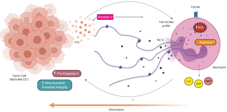
Immune system cells, including neutrophils, are recruited by the tumor microenvironment as a site of chronic inflammation and begin to favor tumor growth. Neutrophils present in the tumor site are called tumor-associated neutrophils (TAN) and can present two phenotypes: N1 (antitumor) or N2 (pro-tumor). Evidence shows the high capacity of immune system cells to interact with extracellular vesicles (Evs) released by tumor cells. Evs can modulate the phenotype of cells within the immune system, contributing to tumor development. Here, we investigated the role of MDA-MB-231-derived Evs upon the polarization of neutrophils towards an N2 phenotype and the underlying mechanisms. We observed that neutrophils treated with Evs released by MDA cells (MDA-Evs) had their half-life increased, increased their chemotactic capacity, and released higher levels of NETs and ROS than neutrophils treated with non-tumoral Evs. We also observed that neutrophils treated with MDA-Evs released increased IL-8, VEGF, MMP9, and increased expression of CD184, an N2-neutrophil marker. Finally, neutrophils treated with MDA-Evs increased tumor cell viability. Our results show that MDA-Evs induce an N2-like phenotype, and the blockage of phosphatidylserine by annexin-V may be an essential agent counter-regulating this effect.

Keywords: breast cancer; extracellular vesicles; inflammation; tumor-associated neutrophils.


Conflict of interest statement

The authors declare that they have no conflict of interest regarding the present study.

Figures

Figure 1
Figure 1
Effect of MDA-EVs on human neutrophils. (A) The nuclear morphology of neutrophils (magnification: 100×) and (B) phosphatidylserine membrane exposure was evaluated for apoptosis using annexin V-FITC and PI binding, respectively. Flow cytometry was used to identify apoptotic cells (annexin-V+/PI– and annexin-V+/PI+). (C) MDA-EVs induce neutrophil chemotaxis compared to MCF10-EVs. (D) The amount of extracellular DNA in the supernatant of neutrophils was quantified using NanoDropTM. The annexin-V concentration was 10 nM. Data are expressed as mean + standard error (SEM). ** p < 0.005; *** p < 0.001. Results are representative of 3–6 independent experiments.
Figure 2
Figure 2
Neutrophils treated with MDA-EVs produce high levels of total ROS and low levels of Nitric Oxide. (A) Neutrophils were incubated for one hour with a CM-H2DCFDA probe, and intracellular total ROS production was monitored using a plate reader. (B) Neutrophils were incubated with a DAF-FMDA probe, and NO production was analyzed using a plate reader. The annexin-V concentration was 10 nM. Data are expressed as mean + standard error (SEM). *** p < 0.001. Results are representative of 8–22 independent experiments.
Figure 3
Figure 3
MDA-EVs induce neutrophils to N2-like phenotype. Neutrophils were polarized for three hours, washed, and conditioned media were collected after two hours. The release of (A) IL-8, (B) VEGF, (C) CCL2, and (D) CCL5 was evaluated by ELISA. (E) Neutrophils were lysed, and the protein extracts were separated using SDS-PAGE with 12% acrylamide gels to identify arginase-1 expression. In the representative image, contrast and brightness were adjusted to better visualize the bands. (F) The supernatant from polarized neutrophils was subjected to electrophoresis using SDS-page with 7.5% acrylamide and gelatin. The gels were then stained with a 30% methanol/10% acetic acid solution containing 0.5% Coomassie blue, and discolored using the same solution without dye. Areas of enzymatic activity appeared as clear bands over the dark background. (G) Neutrophils were incubated for 15 min with FITC-conjugated anti-CD95 (H) and APC-conjugated anti-CD184, following which these protein contents were assessed using flow cytometry. The annexin-V concentration was 10 nM. Data are expressed as mean + standard error (SEM). * p ≤ 0.05; ** p < 0.005; *** p < 0.001. Results are representative of 4–15 independent experiments.
Figure 4
Figure 4
N2-like neutrophils increase tumor cell viability. (A) Polarized neutrophils were co-cultured with previously seeded MDA-MB-231 cells (1:10 ratio of MDA: neutrophils) for 24 h. Cell suspensions were incubated with MTT (5 mg/mL) during the last four hours, and post this, MTT metabolization by viable MDA cells was monitored using a plate reader at 570 nm. (B) MDA-MB-231 cells were primed by N1 or N2-like neutrophils, using a transwell insert. After 24 h, the tumor cells were lysed, and the protein extracts were separated using SDS-PAGE with 12% acrylamide gels to identify pro-caspase expression. The annexin-V concentration was 10 nM. Data are expressed as mean + standard error (SEM). (C) MDA-MB-231 cells were primed by N1 or N2-like neutrophils, using a transwell insert. After 24 h, the mitochondrial membrane potential was monitored using the JC-1 probe. * p ≤ 0.05; ** p < 0.005; *** p ≤ 0.001; **** p ≤ 0.0001 Results are representative of 3–5 independent experiments.
Figure 5
Figure 5
Proposed model for N2-like neutrophil polarization by EVs derived from breast tumor cells (MDA-MB-231). Tumor cells produce and release EVs, which create a chemoattractant environment for neutrophils, protect neutrophils from spontaneous apoptosis, induce the release of NETs and production of total intracellular ROS. Furthermore, neutrophils are induced to a pro-tumor N2-like phenotype, exhibiting increased molecular markers such as IL-8, VEGF, MMP-9, arginase-1, and CXCR4. Neutrophils can also increase the viability of breast tumor cells in vitro. Created with Biorender.com accessed on 6 January 2022.

Similar articles

Cited by

References

    1. Ocana A., Nieto-Jiménez C., Pandiella A., Templeton A.J. Neutrophils in cancer: Prognostic role and therapeutic strategies. Mol. Cancer. 2017;16:137. doi: 10.1186/s12943-017-0707-7. - DOI - PMC - PubMed
    1. Shaul M.E., Fridlender Z.G. Cancer-related circulating and tumor-associated neutrophils—Subtypes, sources and function. FEBS J. 2018;285:4316–4342. doi: 10.1111/febs.14524. - DOI - PubMed
    1. Hanahan D., Weinberg R.A. Hallmarks of cancer: The next generation. Cell. 2011;144:646–674. doi: 10.1016/j.cell.2011.02.013. - DOI - PubMed
    1. Kim J., Bae J.-S. Tumor-Associated Macrophages and Neutrophils in Tumor Microenvironment. Mediat. Inflamm. 2016;2016:6058147. doi: 10.1155/2016/6058147. - DOI - PMC - PubMed
    1. Mantovani A., Marchesi F., Jaillon S., Garlanda C., Allavena P. Tumor-associated myeloid cells: Diversity and therapeutic targeting. Cell. Mol. Immunol. 2021;18:566–578. doi: 10.1038/s41423-020-00613-4. - DOI - PMC - PubMed

Publication types