Review
. 2022 Jun 11;11(12):1895.
doi: 10.3390/cells11121895.

HypoxaMIRs: Key Regulators of Hallmarks of Colorectal Cancer

Affiliations
Review

HypoxaMIRs: Key Regulators of Hallmarks of Colorectal Cancer

Jossimar Coronel-Hernández et al. Cells. .

Abstract

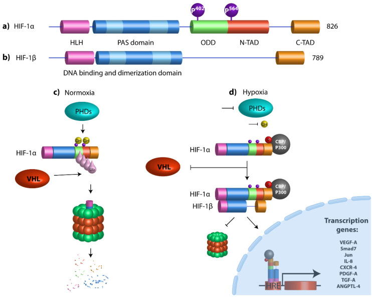
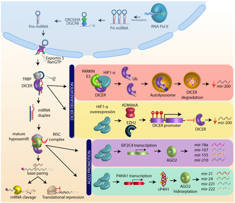
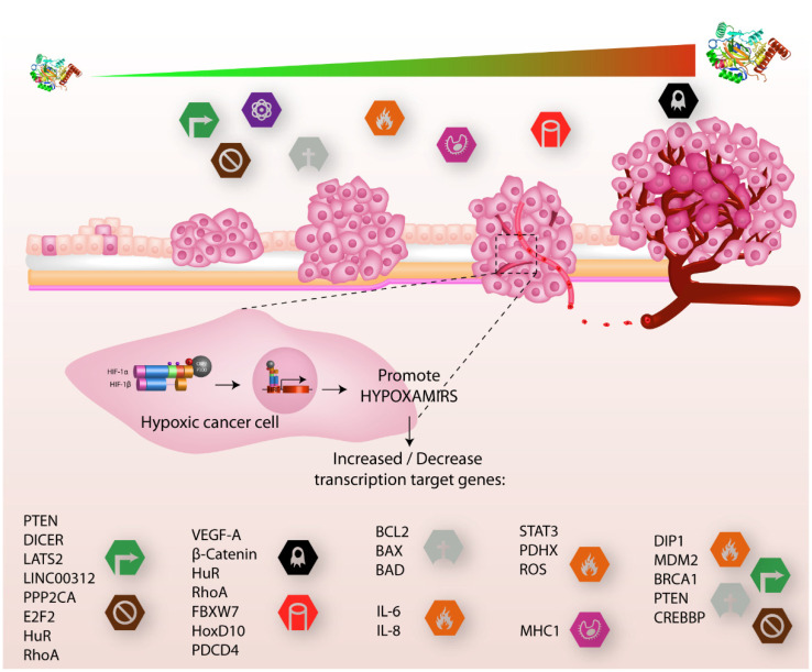
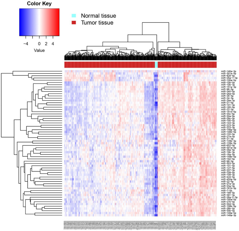
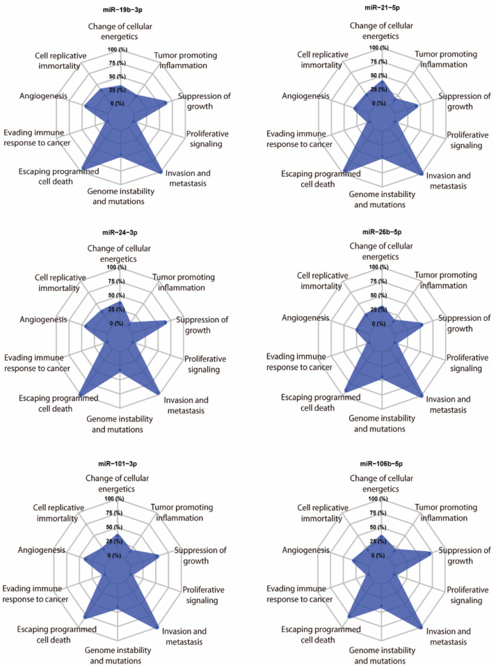
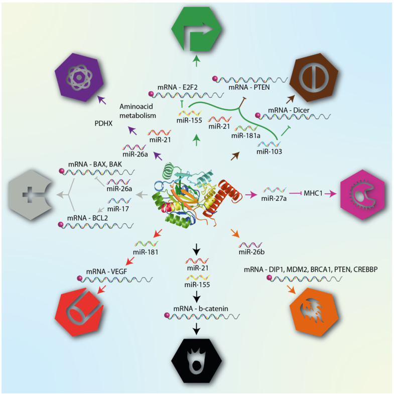
Hypoxia in cancer is a thoroughly studied phenomenon, and the logical cause of the reduction in oxygen tension is tumor growth itself. While sustained hypoxia leads to death by necrosis in cells, there is an exquisitely regulated mechanism that rescues hypoxic cells from their fatal fate. The accumulation in the cytoplasm of the transcription factor HIF-1α, which, under normoxic conditions, is marked for degradation by a group of oxygen-sensing proteins known as prolyl hydroxylases (PHDs) in association with the von Hippel-Lindau anti-oncogene (VHL) is critical for the cell, as it regulates different mechanisms through the genes it induces. A group of microRNAs whose expression is regulated by HIF, collectively called hypoxaMIRs, have been recognized. In this review, we deal with the hypoxaMIRs that have been shown to be expressed in colorectal cancer. Subsequently, using data mining, we analyze a panel of hypoxaMIRs expressed in both normal and tumor tissues obtained from TCGA. Finally, we assess the impact of these hypoxaMIRs on cancer hallmarks through their target genes.

Keywords: HIF-1α; angiogenesis; hypoxaMIR; metastasis; miRNA network; miRNA regulation; microRNAs; transcription factor.


Conflict of interest statement

The authors declare no conflict of interest.

Figures

Figure 1
Figure 1
Structural domains of HIF-1α and HIF-1β and schematic diagram of HIF pathway. (a) HIF-1α contains a basic helix–loop–helix (HLH) system and two dimerization domains (PAS-A and PAS-B) that mediate DNA binding and dimerization, respectively. An oxygen-dependent degradation domain (ODD) is required for oxygen-dependent hydroxylation and degradation under normoxia conditions. The transactivation domains (N-and C-Terminal TADs) are both responsible for the transcriptional activity. N-TAD domain is located within the ODD domain and C-TAD domain at the C-terminal region of the protein. Prolyl hydroxylases (PHDs) hydroxylate HIF-1α proline residues 402 and 564. (b) HIF-1β is the aryl hydrocarbon receptor nuclear translocator (ARNT), which has a bHLH domain, PAS-A and PAS-B domains, and only one C-terminal transactivation domain. (c) Under normoxic conditions, HIF-1α undergoes hydroxylation by prolyl-4-hydroxylases (PHDs) at proline residues 402 and 564, which promotes the destabilization of the HIF-1α protein, which allows the von Hippel-Lindau (VHL) protein to bind it, resulting in polyubiquitination, leading to proteasome-mediated degradation. (d) Under hypoxic conditions, PHDs are inactivated, promoting HIF-1α stabilization. HIF-1α dimerizes with its partner HIF-1β (ARNT), they are translocated to the nucleus, and, in combination with the transcriptional coactivator CBP/P300, they bind to genomic DNA at hypoxia response elements (HREs) to activate the transcription of target genes.
Figure 2
Figure 2
MicroRNA biogenesis and HIF-1α role. MicroRNA (miRNA) are transcribed as primary miRNAs (pri-miRNAs) by RNA polymerase II (Pol II) in the nucleus. The pri-miRNAs are cleaved by the endonuclease DROSHA/DGCR8 (DiGeorge syndrome critical region 8) to a shorter pre-miRNA hairpin structure (60–70 nucleotides). The pre-miRNAs are exported from the nucleus to the cytoplasm by exportin-5-Ran-GTP (XPO5), and are processed by DICER1/TRBP, a ribonuclease III (RIII) enzyme that produces miRNA duplex (~21-nucleotide). The final step of miRNA maturation is the selective functional strand of small RNA duplex into RNA-induced silencing complex (RISC), which includes DICER, TRPB, and Argonaut (AGO). One strand of the mature miRNA (the guide strand) is loaded with Argonaut (AGO2) to form a miRNA-induced silencing complex (miRISC) that targets mRNAs by a complementary sequence leading to mRNA degradation and translational repression. HIF-1α negatively regulates DICER through autolysosome degradation and promoter methylation, whereas HIF1-α positively regulates AGO by target transcription and promotes AGO hydroxilation (see text).
Figure 3
Figure 3
HIF-α involved in cancer progression. In the early stages of tumor growth, cells acquire distinctive hallmarks that promote cancer progression, such as sustaining proliferative signals, evasion growth suppression, reprogramming energy metabolism, and resistance to cell death, while in the final stages of tumor growth, hallmarks which are added include inflammation, avoiding immune destruction, and, finally, metastasis and angiogenesis. These features are regulated by different genes (lower part) that are activated or inhibited by different hypoxaMIRs, which are regulated positively and negatively by the transcription factor HIF1-α, which, in turn, increases the concentration of proteins as tumor progression increases.
Figure 4
Figure 4
Heat map of differential expression of hypoxaMIRs in human normal and tumor tissues. Differential expression of hypoxaMIRs was identified in normal tissue and tumor tissue by a miRNA body map along with the hierarchical cluster analysis. Expression of hypoxaMIRs is represented as blue (downregulated), red (upregulated), and white colors (no significant change or absence of data).
Figure 5
Figure 5
In silico analysis. Radar chart of the relevant hallmarks of cancer simultaneously modulated by the selected hypoxaMIRs after analysis. The levels show the proportion of the number of putative gene targets for each class.
Figure 6
Figure 6
HypoxaMIRs associated with their corresponding cancer hallmark. The figure lists some hypoxaMIRs (previously described in this review) that regulate genes involved in the different hallmarks of cancer, as defined by Hanahan and Weinberg. These hallmarks are cellular mechanisms acquired during tumor transformation, and include proliferation, apoptosis, angiogenesis, invasion, and metastasis.

Similar articles

Cited by

References

    1. Sung H., Ferlay J., Siegel R.L., Laversanne M., Soerjomataram I., Jemal A., Bray F. Global Cancer Statistics 2020: GLOBOCAN Estimates of Incidence and Mortality Worldwide for 36 Cancers in 185 Countries. CA Cancer J. Clin. 2021;71:209–249. doi: 10.3322/caac.21660. - DOI - PubMed
    1. Pritchard C.C., Grady W.M. Colorectal cancer molecular biology moves into clinical practice. Gut. 2011;60:116–129. doi: 10.1136/gut.2009.206250. - DOI - PMC - PubMed
    1. Yang L., Belaguli N., Berger D.H. MicroRNA and Colorectal Cancer. World J. Surg. 2009;33:638–646. doi: 10.1007/s00268-008-9865-5. - DOI - PubMed
    1. Banerjee N., Kim H., Talcott S., Mertens-Talcott S. Pomegranate polyphenolics suppressed azoxymethane-induced colorectal aberrant crypt foci and inflammation: Possible role of miR-126/VCAM-1 and miR-126/PI3K/AKT/mTOR. Carcinogenesis. 2013;34:2814–2822. doi: 10.1093/carcin/bgt295. - DOI - PubMed
    1. Pan Z., Tian Y., Niu G., Cao C. Role of microRNAs in remodeling the tumor microenvironment (Review) Int. J. Oncol. 2020;56:407. doi: 10.3892/ijo.2019.4952. - DOI - PMC - PubMed

Publication types

Substances