. 2022 Jun 11;11(12):1899.
doi: 10.3390/cells11121899.

Alpha-Keratin, Keratin-Associated Proteins and Transglutaminase 1 Are Present in the Ortho- and Parakeratinized Epithelium of the Avian Tongue

Affiliations

Alpha-Keratin, Keratin-Associated Proteins and Transglutaminase 1 Are Present in the Ortho- and Parakeratinized Epithelium of the Avian Tongue

Kinga Skieresz-Szewczyk et al. Cells. .

Abstract

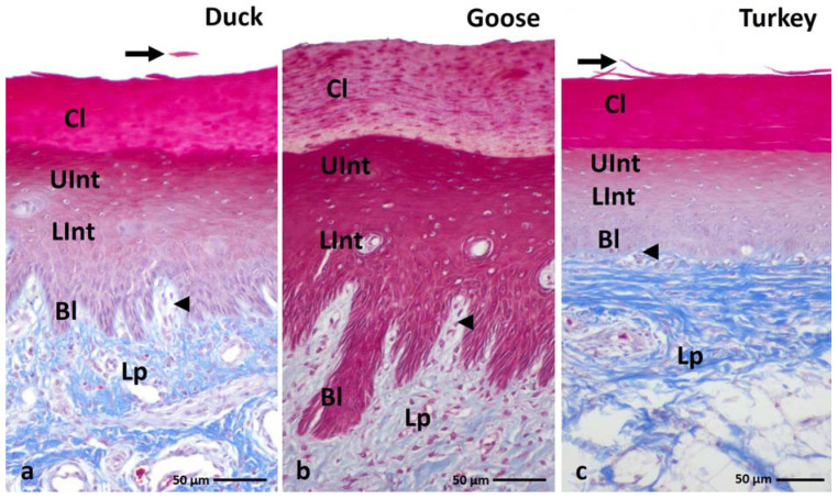
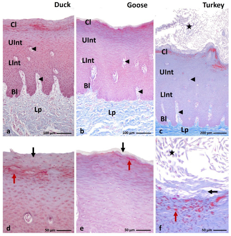
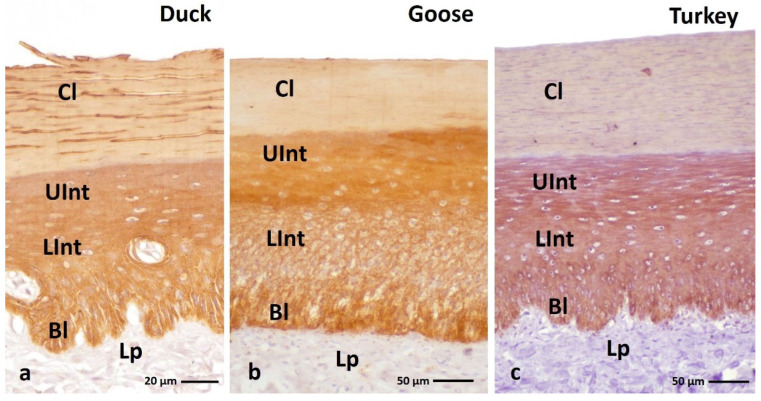
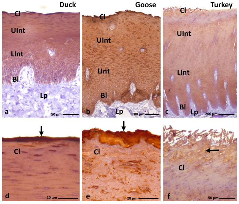
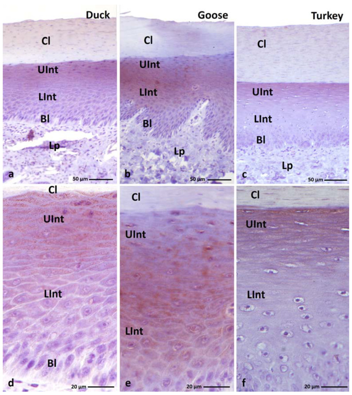
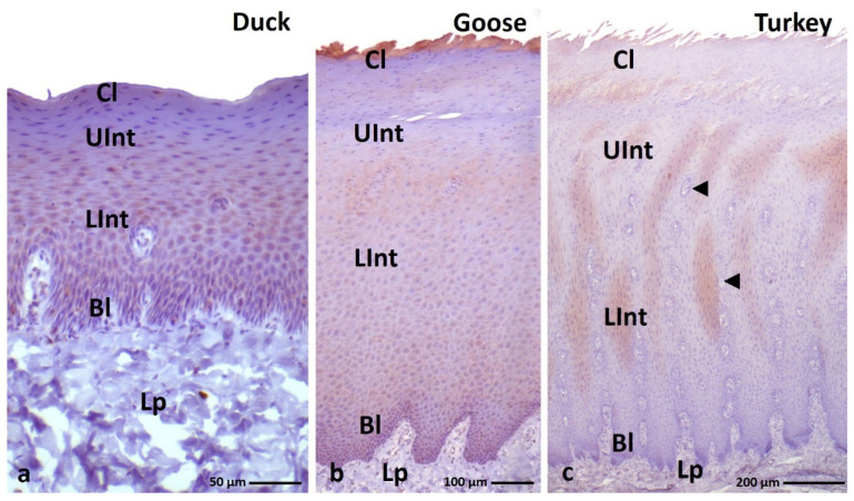
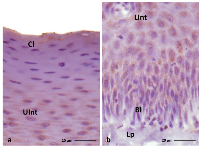
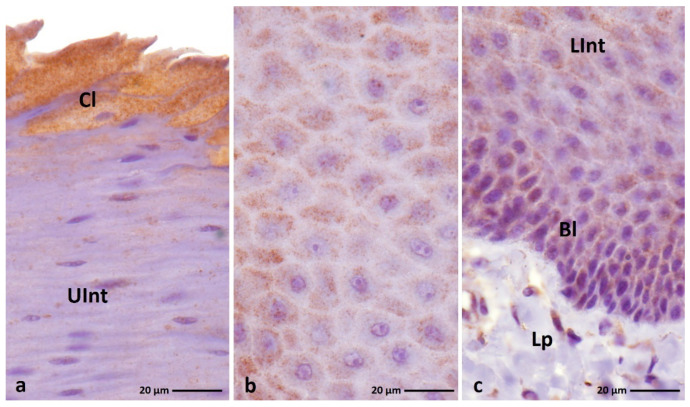
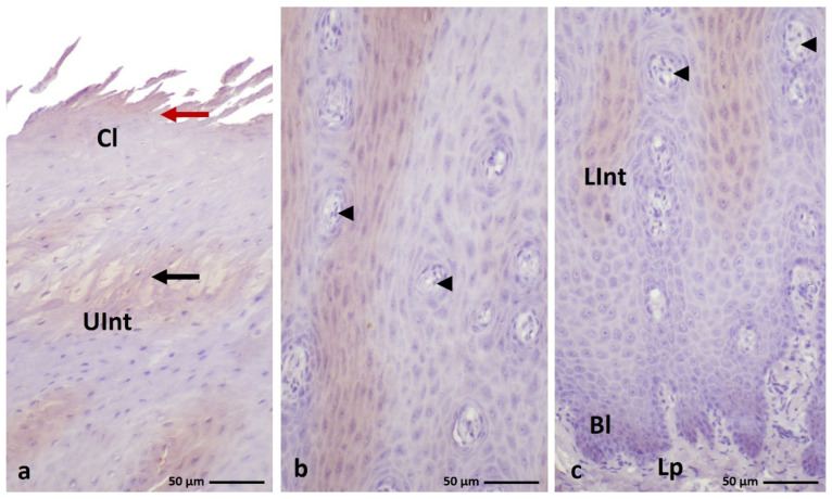
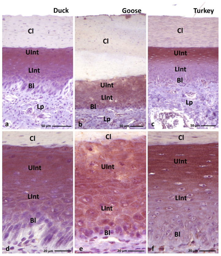
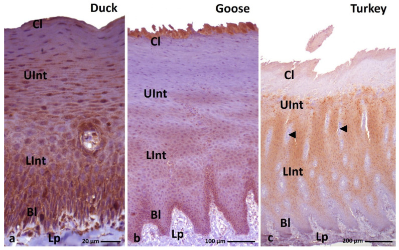
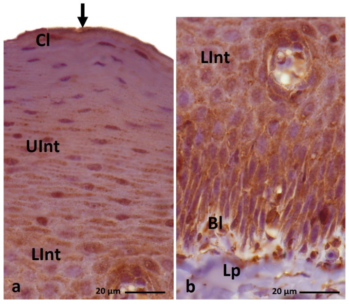
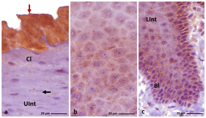
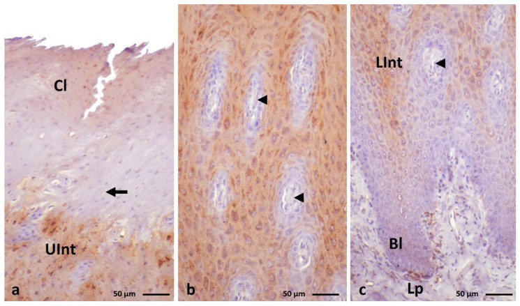
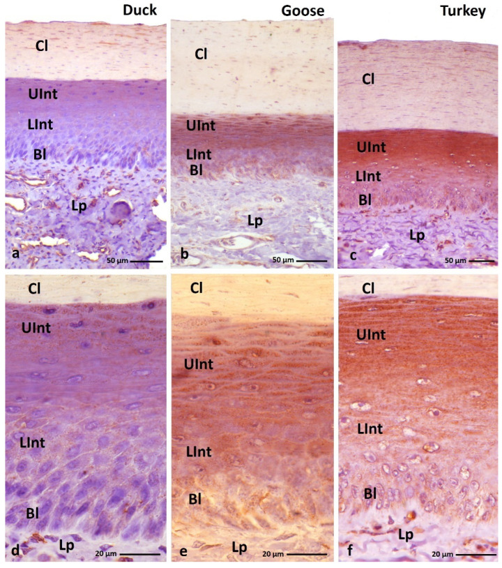
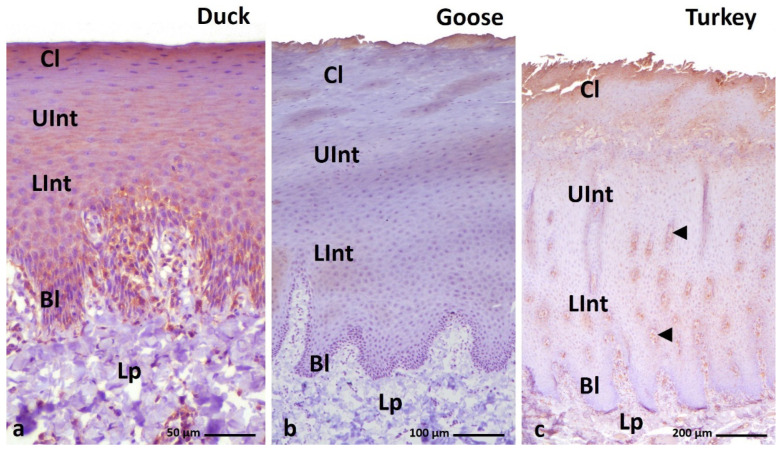
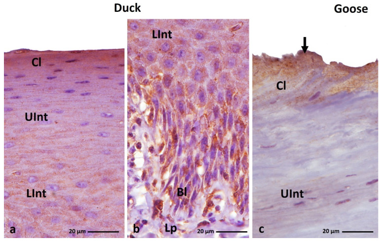
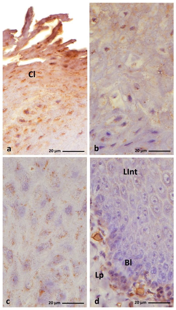
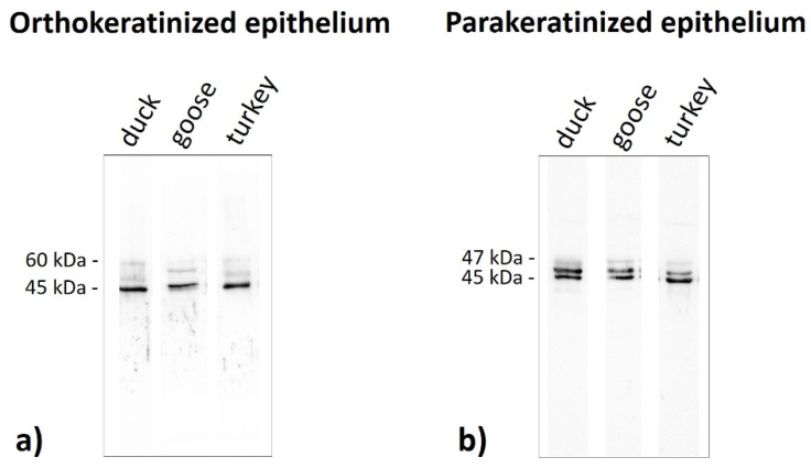
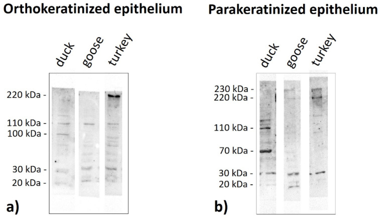
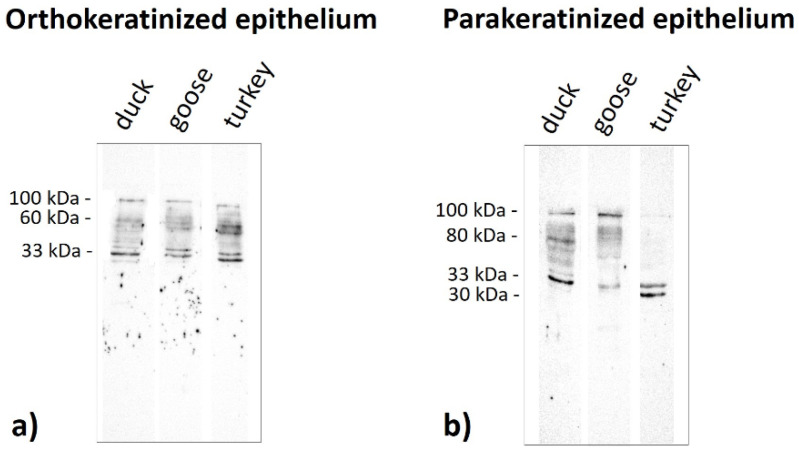
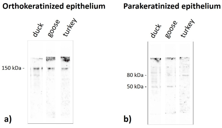
The lingual mucosa in birds is covered with two specific types of multilayered epithelia, i.e., the para- and orthokeratinized epithelium, that differ structurally and functionally. Comprehensive information on proteins synthesized in keratinocyte during their cytodifferentiation in subsequent layers of multilayered epithelia in birds concerns only the epidermis and are missing the epithelia of the lingual mucosa. The aim of the present study was to perform an immunohistochemical (IHC) and molecular analysis (WB) of bird-specific alpha-keratin, keratin-associated proteins (KAPs), namely filaggrin and loricrin, as well as transglutaminase 1 in the para- and orthokeratinized epithelium covering the tongue in the domestic duck, goose, and turkey. The results reveal the presence of alpha-keratin and KAPs in both epithelia, which is a sign of the cornification process. In contrast to the epidermis, the main KAPs involved in the cornification process of the lingual epithelia in birds is loricrin. Stronger expression with KAPs and transglutaminase 1 in the orthokeratinized epithelium than in the parakeratinized epithelium may determine the formation of a more efficient protective mechanical barrier. The presence of alpha-keratin, KAPs, and transglutaminase 1 epitopes characteristic of epidermal cornification in both types of the lingual epithelia may prove that they are of ectodermal origin.

Keywords: KAPs; alpha-keratin; birds; cornification; orthokeratinized epithelium; parakeratinized epithelium; tongue.


Conflict of interest statement

The authors declare no conflict of interest.

Figures

Figure 1
Figure 1
(ac) Cross-section of the orthokeratinized epithelium. Black arrows show exfoliated superficial cells of the cornified layer. Arrowheads point to the connective tissue papillae. Bl—basal layer, Lp—lamina propria, LInt—the lower part of the intermediate layer, UInt—upper part of the intermediate layer, Cl—cornified layer. Masson–Goldner staining.
Figure 2
Figure 2
(ac) Cross-section of the parakeratinized epithelium. Arrowheads point to the connective tissue papillae. Asterisks show massively exfoliated superficial cells of the cornified layer. (df) Higher magnification of the cornified layer of the parakeratinized epithelium. Black arrows show superficial cells of the cornified layer, which cytoplasm stained with pink or light pink color. Red arrows point to cells with red-colored cell cytoplasm of the cornified layer. Bl—basal layer, Lp—lamina propria, LInt—the lower part of the intermediate layer, UInt—upper part of the intermediate layer, Cl—cornified layer. Masson–Goldner staining.
Figure 3
Figure 3
(ac) IHC staining of the alpha-keratin in the orthokeratinized epithelium. Bl—basal layer, Lp—lamina propria, Lint—the lower part of the intermediate layer, UInt—upper part of the intermediate layer, Cl—cornified layer. Counterstaining with Mayer’s hematoxylin.
Figure 4
Figure 4
(ac) IHC staining of the alpha-keratin in the parakeratinized epithelium. (df) Higher magnification of the cornified layer. Arrows point to the superficial cells of the cornified layer with a strong staining reaction. Bl—basal layer, Lp—lamina propria, LInt—lower part of the intermediate layer, UInt—upper part of the intermediate layer, Cl—cornified layer. Counterstaining with Mayer’s hematoxylin.
Figure 5
Figure 5
(ac) IHC staining of the filaggrin in the orthokeratinized epithelium. (df) Higher magnification of basal, intermediate, and cornified layers of the orthokeratinized epithelium. Bl—basal layer. Lp—lamina propria, LInt—the lower part of the intermediate layer, UInt—upper part of the intermediate layer, Cl—cornified layer. Counterstaining with Mayer’s hematoxylin.
Figure 6
Figure 6
(ac) IHC staining of the filaggrin in the parakeratinized epithelium. Arrowheads point to the connective tissue papillae. Bl—basal layer, Lp—lamina propria, LInt—the lower part of the intermediate layer, UInt—upper part of the intermediate layer, Cl—cornified layer. Counterstaining with Mayer’s hematoxylin.
Figure 7
Figure 7
IHC staining of the filaggrin in the parakeratinized epithelium in duck. Higher magnification of (a) the upper part of the intermediate layer and cornified layer and (b) the basal layer and lower part of the intermediate layer. Bl—basal layer, Lp—lamina propria, LInt—lower part of the intermediate layer, UInt—upper part of the intermediate layer, Cl—cornified layer. Counterstaining with Mayer’s hematoxylin.
Figure 8
Figure 8
IHC staining of the filaggrin in the parakeratinized epithelium in goose. Higher magnification of (a) the upper part of the intermediate layer and cornified layer, (b) the lower part of the intermediate layer, and (c) the lower part of the intermediate layer and basal layer. Bl—basal layer, Lp—lamina propria, LInt—lower part of the intermediate layer, UInt—upper part of the intermediate layer, Cl—cornified layer. Counterstaining with Mayer’s hematoxylin.
Figure 9
Figure 9
IHC staining of the filaggrin in the parakeratinized epithelium in the turkey. Higher magnification of (a) the upper part of the intermediate layer and cornified layer, (b) the lower part of the intermediate layer, and (c) the lower part of the intermediate layer and basal layer. The black arrow points to the 2–3 cell layers of the upper part of the intermediate layer with a weak staining reaction. The red arrow shows the superficial cells of the cornified layer with a weak staining reaction. Arrowheads point to the connective tissue papillae. Bl—basal layer, Lp—lamina propria, LInt—lower part of the intermediate layer, UInt—upper part of the intermediate layer, Cl—cornified layer. Counterstaining with Mayer’s hematoxylin.
Figure 10
Figure 10
(ac) IHC staining of the loricrin in the orthokeratinized epithelium. (d–f) Higher magnification of basal, intermediate, and cornified layers of the orthokeratinized epithelium. Bl—basal layer, Lp—lamina propria, LInt—lower part of the intermediate layer, UInt—upper part of the intermediate layer, Cl—cornified layer. Counterstaining with Mayer’s hematoxylin.
Figure 11
Figure 11
(ac) IHC staining of the loricrin in the parakeratinized epithelium. Arrowheads point to the connective tissue papillae. Bl—basal layer, Lp—lamina propria, LInt—the lower part of the intermediate layer, UInt—upper part of the intermediate layer, Cl—cornified layer. Counterstaining with Mayer’s hematoxylin.
Figure 12
Figure 12
IHC staining of the loricrin in the parakeratinized epithelium in the duck. Higher magnification of (a) the intermediate layer and cornified layer and (b) the lower part of the intermediate layer and basal layer. Arrow shows the superficial cell with a strong staining reaction. Bl—basal layer, Lp—lamina propria, LInt—lower part of the intermediate layer, UInt—upper part of the intermediate layer, Cl—cornified layer. Counterstaining with Mayer’s hematoxylin.
Figure 13
Figure 13
IHC staining of the loricrin in the parakeratinized epithelium in the goose. Higher magnification of (a) the upper part of the intermediate layer and cornified layer, (b) the lower part of the intermediate layer, and (c) the basal layer and lower part of the intermediate layer. Red arrow points to the superficial cell with a strong staining reaction. Black arrow points to the cell of the upper part of the intermediate layer with a weak staining reaction. Bl—basal layer, Lp—lamina propria, LInt—lower part of the intermediate layer, UInt—upper part of the intermediate layer, Cl—cornified layer. Counterstaining with Mayer’s hematoxylin.
Figure 14
Figure 14
IHC staining of the loricrin in the parakeratinized epithelium in the turkey. Higher magnification of (a) the upper part of the intermediate layer and cornified layer, (b) the lower part of the intermediate layer, and (c) the basal layer and lower part of the intermediate layer. The black arrow points to the cells of the intermediate layer with weak staining reaction. Arrowheads point to the connective tissue papillae. Bl—basal layer, Lp—lamina propria, LInt—the lower part of the intermediate layer, UInt—upper part of the intermediate layer, Cl—cornified layer. Counterstaining with Mayer’s hematoxylin.
Figure 15
Figure 15
(ac) IHC staining of the TGM-1 in the orthokeratinized epithelium. (df) Higher magnification of basal, intermediate, and cornified layers of the orthokeratinized epithelium. Bl—basal layer, Lp—lamina propria, LInt—the lower part of the intermediate layer, UInt—upper part of the intermediate layer, Cl—cornified layer. Counterstaining with Mayer’s hematoxylin.
Figure 16
Figure 16
(ac) IHC staining of the TGM-1 in the parakeratinized epithelium. Arrowheads point to the connective tissue papillae. Bl—basal layer, Lp—lamina propria, LInt—the lower part of the intermediate layer, UInt—upper part of the intermediate layer, Cl—cornified layer. Counterstaining with Mayer’s hematoxylin.
Figure 17
Figure 17
IHC staining of the TGM-1 in the parakeratinized epithelium. Higher magnification of (a) the upper part of the intermediate layer and cornified layer in the duck, (b) the basal layer and lower part of the intermediate layer in the duck, and (c) the upper part of the intermediate layer and cornified layer in the goose. The black arrow points to the superficial cells of the cornified layer with a medium staining reaction. Bl—basal layer, Lp—lamina propria, LInt—lower part of the intermediate layer, UInt—upper part of the intermediate layer, Cl—cornified layer. Counterstaining with Mayer’s hematoxylin.
Figure 18
Figure 18
IHC staining of the TGM-1 in the parakeratinized epithelium in the turkey. Higher magnification of (a) the cornified layer, (b) the upper part of the intermediate layer, (c) the lower part of the intermediate layer, and (d) the basal layer and lower part of the intermediate layer. Bl—basal layer, Lp—lamina propria, LInt—the lower part of the intermediate layer, UInt—upper part of the intermediate layer, Cl—cornified layer. Counterstaining with Mayer’s hematoxylin.
Figure 19
Figure 19
Immunoblots of the (a) orthokeratinized and (b) parakeratinized epithelium with the alpha-keratin antiserum in the duck, goose, and turkey.
Figure 20
Figure 20
Immunoblots of the (a) orthokeratinized and (b) parakeratinized epithelium with the filaggrin antiserum in the duck, goose, and turkey.
Figure 21
Figure 21
Immunoblots of the (a) orthokeratinized and (b) parakeratinized epithelium with the loricrin antiserum in the duck, goose, and turkey.
Figure 22
Figure 22
Immunoblots of the (a) orthokeratinized and (b) parakeratinized epithelium with the TGM-1 in the duck, goose, and turkey.

Similar articles

Cited by

References

    1. Alibardi L. Adaptation to the land: The skin of reptiles in comparison to that of amphibians and endoderm amniotes. J. Exp. Zool. B Mol. Dev. Evol. 2003;29:12–41. doi: 10.1002/jez.b.24. - DOI - PubMed
    1. Alibardi L. Structural and Immunocytochemical Characterization of Keratinization in Vertebrate Epidermis and Epidermal Derivatives. Int. Rev. Cytol. 2006;253:177–259. doi: 10.1016/s0074-7696(06)53005-0. - DOI - PubMed
    1. Bragulla H.H., Homberger D.G. Structure and functions of keratin proteins in simple, stratified, keratinized and cornified epithelia. J. Anat. 2009;214:516–559. doi: 10.1111/j.1469-7580.2009.01066.x. - DOI - PMC - PubMed
    1. Giroud A., Leblond C.P. The keratinization of epidermis and its derivates. Especially the hair, as shown by X-ray diffraction and histochemical studies. Ann. N. Y. Acad. Sci. 1951;53:613–626. doi: 10.1111/j.1749-6632.1951.tb31963.x. - DOI - PubMed
    1. Rhodin J.A.G., Reith E.J. Ultrastructure of keratin in oral mucosa, skin, esophagus, claw and hair. In: Butcher E.O., Signnaes R.F., editors. Fundamentals of Keratinization. American Association for the Advancement of Science; Washington, DC, USA: pp. 61–94.

Publication types

LinkOut - more resources