. 2022 Jun 12;11(12):1908.
doi: 10.3390/cells11121908.

Super-Resolution Imaging of Fas/CD95 Reorganization Induced by Membrane-Bound Fas Ligand Reveals Nanoscale Clustering Upstream of FADD Recruitment

Affiliations

Super-Resolution Imaging of Fas/CD95 Reorganization Induced by Membrane-Bound Fas Ligand Reveals Nanoscale Clustering Upstream of FADD Recruitment

Nicholas Frazzette et al. Cells. .

Abstract

Signaling through the TNF-family receptor Fas/CD95 can trigger apoptosis or non-apoptotic cellular responses and is essential for protection from autoimmunity. Receptor clustering has been observed following interaction with Fas ligand (FasL), but the stoichiometry of Fas, particularly when triggered by membrane-bound FasL, the only form of FasL competent at inducing programmed cell death, is not known. Here we used super-resolution microscopy to study the behavior of single molecules of Fas/CD95 on the plasma membrane after interaction of Fas with FasL on planar lipid bilayers. We observed rapid formation of Fas protein superclusters containing more than 20 receptors after interactions with membrane-bound FasL. Fluorescence correlation imaging demonstrated recruitment of FADD dependent on an intact Fas death domain, with lipid raft association playing a secondary role. Flow-cytometric FRET analysis confirmed these results, and also showed that some Fas clustering can occur in the absence of FADD and caspase-8. Point mutations in the Fas death domain associated with autoimmune lymphoproliferative syndrome (ALPS) completely disrupted Fas reorganization and FADD recruitment, confirming structure-based predictions of the critical role that these residues play in Fas-Fas and Fas-FADD interactions. Finally, we showed that induction of apoptosis correlated with the ability to form superclusters and recruit FADD.

Keywords: CD95; Fas; PALM imaging; TNFR superfamily; receptor signaling; super-resolution microscopy.


Conflict of interest statement

R.M.S. is currently an employee of Novartis and owns Novartis stock.

Figures

Figure 1
Figure 1
SIM and PALM imaging of Fas receptor clustering after interaction with SLB-anchored FasL. (A) Schematics of the experimental setup used to image the spatial distribution of Fas receptors following stimulation with Fas ligand (FasL). Jurkat cells expressing Fas-EGFP were adhered to supported lipid bilayers (SLBs) containing FasL, ICAM-1, and anti-TCR (anti-CD3) antibodies. (BD) Spatial distribution of Fas receptors on plasma membrane of Jurkat cells placed on SLBs. (B) Diffraction-limited TIRF images showing the distribution of Fas-EGFP (green) T cell receptors (labeled with anti-CD3 antibodies, red), with (bottom panel) or without (top panel) FasL added to the SLB. Scale bar = 2 microns. (C) PALM images of nanoscale spatial distribution of Fas-PAGFP receptor fusion proteins before (right panel) or after (left panel) engagement with FasL. The relative probability density of Fas receptors is mapped on to each pixel using the density color map on the right. Scale bar = 1 micron. (D) Spatial distribution of protein islands identified by cluster analysis of Fas PALM images acquired before (left) or after (right) stimulation with FasL. Individual receptors within a protein island are color-coded according to the number of Fas receptors (protein island occupancy, ϕisland) present in the protein island. Scale bar = 1 micron. (EG) Plot of physical parameters of the protein islands obtained from spatial cluster analysis of the distribution of Fas receptors in PALM images. (E) Plot of distribution of number of Fas receptors per protein island (ϕisland) with or without stimulation with Fas ligand. Each circle represents a single protein island. Data are from four cells for each condition and are compared by two-tailed Mann–Whitney U test (p < 0.0001). (F) Plot of distribution of total Fas receptors that reside in protein islands containing indicated number of receptors. Data are shown for both with (magenta) or without (green) stimulation with Fas ligand. (G) Plot showing the frequency of occurrence of protein islands containing indicated number of Fas receptors (ϕisland). (H) Plot of percentage of total Fas receptors residing in protein islands of indicated size (radius in nm).
Figure 2
Figure 2
Contributions of structural domains of Fas to nanoscale receptor clustering. (A) Spatial distribution of Fas receptors was quantified following interaction of Jurkat cells with different FasL-containing substrates. FasL was either incorporated into fluid DOPC-based supported lipid bilayers, gel-like DSPC-based supported lipid bilayers, or immobilized onto polylysine-coated coverslips. Cluster parameters were evaluated with or without stimulation with FasL. (Left panel) Plot of distribution of percentage of Fas receptors in protein islands containing indicated number of proteins. (Right panel) Distribution of percentage of proteins residing in protein islands of indicated size (radius in nm). (B) Schematic of Fas receptor showing the location of preligand assembly domain (PLAD), the palmitoylation site CysC199, the death domain, and the ALPS mutations. (C,D) Cluster parameters of protein islands of wild-type Fas or indicated Fas receptor mutants (C: structural mutants, D: ALPS mutants) with or without interactions with FasL. Plots of distribution of percentage of proteins in protein islands of indicated protein occupancy (left panel, C,D), or within protein islands of indicated size (right panel, C,D). (E) Fas-deficient Jurkat cells (RapoC2) were cotransfected with homotypic pairs of indicated receptors, each tagged with either mCerulean3 or mVenus. 24 h post-transfection, cells were treated with FasL-LZ, and FRET between the pairs of Fas molecules was measured at the indicated timepoints. Fold change was calculated as the change in FRET intensity over similarly transfected, but untreated, cells. A minimum of n = 4 was performed for each experimental condition. (F) Wild-type Fas receptors tagged with either mCerulean3 or mVenus were cotransfected into wild-type Fas-sufficient Jurkat cells or mutant Jurkat cells deficient for Fas (RapoC2), FADD (I2.1), or caspase-8 (I9.2). Cells were stimulated with FasL-LZ, as in (E), and assessed for Fas–Fas FRET via flow cytometry on the Cerulean3+Venus+ viable population. Fold change was calculated as in (E). A minimum n = 3 was performed for each experimental condition.
Figure 3
Figure 3
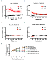
Cross-correlation and FRET analysis of Fas–FADD interactions. (AD) Spatial interactions of FADD with either wild-type Fas or different mutants of Fas receptors on plasma membrane of Jurkat cells were quantified by spatial cross-correlation analysis of multicolor PALM images. PAGFP-tagged receptor molecules and FADD-PAmCh were coexpressed in Jurkat cells and imaged with or without interactions with FasL on DOPC-based fluid supported lipid bilayers. Plots of spatial cross-correlation (c(r)) between FADD-PAmCh and PAGFP-labeled wild-type Fas receptor (A), palmitoylation deficient Fas-C99V (B), Fas-ΔDD lacking death domain (C), or the ALPS mutant Fas-A257D (D). Value of c(r) >1 indicates increased spatial clustering of the two kinds of molecules, c(r) < 1 indicates spatial exclusion, and c(r) = 1 indicates random distribution of the molecules. (E) Fas-deficient RapoC2 Jurkat cells were cotransfected with a Cerulean3-tagged FADD containing only the death domain (FADD DD) and either wild-type or mutant Fas receptors as indicated. Cells were treated with FasL-LZ and changes in FRET between Fas and FADD were analyzed as in Figure 2E,F. A minimum of three independent experiments were performed (n = 3).
Figure 4
Figure 4
Relationship between Fas clustering and apoptosis induction in T cells. (A) Fas-deficient Jurkat cells (RapoC2) were transfected with mVenus-tagged WT or mutant Fas. 24 h post-transfection, cells were treated with increasing amounts of FasL-LZ for 6 h prior to staining with Annexin V, DiIC(5), and Live/Dead, and being analyzed by flow cytometry. (B) Activated CD4+ T cells from mice with the indicated genotype were stained for Fas surface expression and assessed by flow cytometry. Data are representative of n = 3 independent experiments. (C) CD4 T cells were isolated from mice of the indicated genotypes and activated for 72 h with anti-CD3/28. Cells were cultured with supplemental IL-2 prior to treatment with FasL-LZ at the indicated concentrations for 6 h, then stained for assessment of cell death via flow cytometry as in (A). (D) CD3/28 activated CD4 T cells from mice of the indicated genotypes were rested in IL-2 and subsequently restimulated with plate-bound anti-CD3 antibody at the indicated concentrations for 16 h prior to assessment for death via flow cytometry as in (A). Panels (AC) are cumulative data from a minimum of three independent experiments (n = 3).

Similar articles

Cited by

References

    1. Wang L., Yang J.K., Kabaleeswaran V., Rice A.J., Cruz A.C., Park A.Y., Yin Q., Damko E., Jang S.B., Raunser S., et al. The Fas-FADD death domain complex structure reveals the basis of DISC assembly and disease mutations. Nat. Struct. Mol. Biol. 2010;17:1324–1329. doi: 10.1038/nsmb.1920. - DOI - PMC - PubMed
    1. Siegel R.M., Chan F.K., Chun H.J., Lenardo M.J. The multifaceted role of Fas signaling in immune cell homeostasis and autoimmunity. Nat. Immunol. 2000;1:469–474. doi: 10.1038/82712. - DOI - PubMed
    1. Peter M.E., Krammer P.H. The CD95(APO-1/Fas) DISC and beyond. Cell Death Differ. 2003;10:26–35. doi: 10.1038/sj.cdd.4401186. - DOI - PubMed
    1. Stranges P.B., Watson J., Cooper C.J., Choisy-Rossi C.M., Stonebraker A.C., Beighton R.A., Hartig H., Sundberg J.P., Servick S., Kaufmann G., et al. Elimination of antigen-presenting cells and autoreactive T cells by Fas contributes to prevention of autoimmunity. Immunity. 2007;26:629–641. doi: 10.1016/j.immuni.2007.03.016. - DOI - PMC - PubMed
    1. Strasser A., Jost P.J., Nagata S. The many roles of FAS receptor signaling in the immune system. Immunity. 2009;30:180–192. doi: 10.1016/j.immuni.2009.01.001. - DOI - PMC - PubMed

Publication types

MeSH terms

Substances