

Computerized Texture Analysis of Optical Coherence Tomography Angiography of Choriocapillaris in Normal Eyes of Young and Healthy Subjects
- PMID: 35741063
- PMCID: PMC9221889
- DOI: 10.3390/cells11121934
Computerized Texture Analysis of Optical Coherence Tomography Angiography of Choriocapillaris in Normal Eyes of Young and Healthy Subjects
Abstract
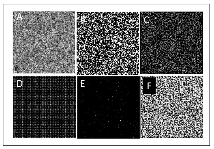
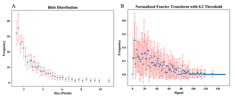
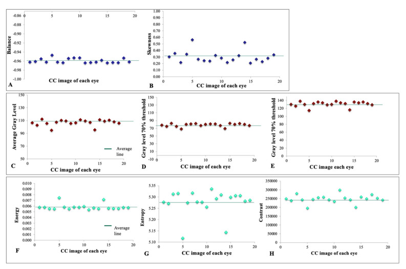
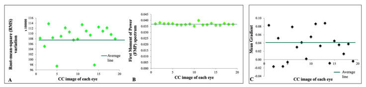
Computerized texture analysis uses higher-order mathematics to identify patterns beyond what the naked eye can recognize. We tested its feasibility in optical coherence tomography angiography imaging of choriocapillaris. Our objective was to determine sets of parameters that provide coherent and consistent output when applied to a homogeneous, healthy group of patients. This observational cross-sectional study involved 19 eyes of 10 young and healthy Caucasian subjects. En-face macular optical coherence tomography angiography of superficial choriocapillaris was obtained by the RTVue-XR Avanti system. Various algorithms were used to extract texture features. The mean and standard deviation were used to assess the distribution and dispersion of data points in each metric among eyes, which included: average gray level, gray level yielding 70% threshold and 30% threshold, balance, skewness, energy, entropy, contrast, edge mean gradient, root-mean-square variation, and first moment of power spectrum, which was compared between images, showing a highly concordant homology between all eyes of participants. We conclude that computerized texture analysis for en-face optical coherence tomography angiography images of choriocapillaris is feasible and provides values that are coherent and tightly distributed around the mean in a homogenous, healthy group of patients. Homology of blob size among subjects may represent a "repeat pattern" in signal density and thus a perfusion in the superficial choriocapillaris of healthy young individuals of the same ethnic background.
Keywords: choriocapillaris; computerized texture analysis; optical coherence tomography angiography.
Conflict of interest statement
The authors declare no conflict of interest. The funders had no role in the design of the study; in the collection, analyses, or interpretation of data; in the writing of the manuscript, or in the decision to publish the results.
Figures
References
-
- Lutty G., Grunwald J., Majji A.B., Uyama M., Yoneya S. Changes in choriocapillaris and retinal pigment epithelium in age-related macular degeneration. Mol. Vis. 1999;5:35. - PubMed
-
- McLeod D.S., Taomoto M., Otsuji T., Green W.R., Sunness J.S., Lutty G.A. Quantifying changes in RPE and choroidal vasculature in eyes with age-related macular degeneration. Investig. Ophthalmol. Vis. Sci. 2002;43:1986–1993. - PubMed
Publication types
MeSH terms
LinkOut - more resources
Full Text Sources
Research Materials
