Review
. 2022 Jun 15;11(12):1935.
doi: 10.3390/cells11121935.

Regulation and Dysregulation of Endothelial Permeability during Systemic Inflammation

Affiliations
Review

Regulation and Dysregulation of Endothelial Permeability during Systemic Inflammation

Katharina E M Hellenthal et al. Cells. .

Abstract

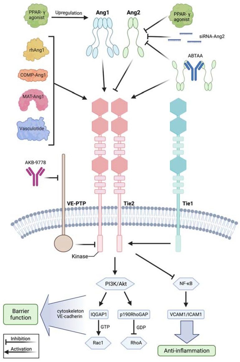
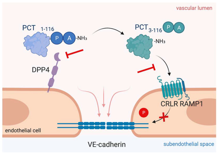
Systemic inflammation can be triggered by infection, surgery, trauma or burns. During systemic inflammation, an overshooting immune response induces tissue damage resulting in organ dysfunction and mortality. Endothelial cells make up the inner lining of all blood vessels and are critically involved in maintaining organ integrity by regulating tissue perfusion. Permeability of the endothelial monolayer is strictly controlled and highly organ-specific, forming continuous, fenestrated and discontinuous capillaries that orchestrate the extravasation of fluids, proteins and solutes to maintain organ homeostasis. In the physiological state, the endothelial barrier is maintained by the glycocalyx, extracellular matrix and intercellular junctions including adherens and tight junctions. As endothelial cells are constantly sensing and responding to the extracellular environment, their activation by inflammatory stimuli promotes a loss of endothelial barrier function, which has been identified as a hallmark of systemic inflammation, leading to tissue edema formation and hypotension and thus, is a key contributor to lethal outcomes. In this review, we provide a comprehensive summary of the major players, such as the angiopoietin-Tie2 signaling axis, adrenomedullin and vascular endothelial (VE-) cadherin, that substantially contribute to the regulation and dysregulation of endothelial permeability during systemic inflammation and elucidate treatment strategies targeting the preservation of vascular integrity.

Keywords: adrenomedullin; angiopoietin-Tie2; capillary leakage; endothelium; procalcitonin; systemic inflammation; vascular permeability.


Conflict of interest statement

The authors declare no conflict of interest.

Figures

Figure 1
Figure 1
Leukocyte extravasation is independent from vascular leakage. Left: Endothelial barrier function is tightly controlled by intercellular junctions including adherens junctions and tight junctions that connect neighboring endothelial cells. Middle: Leukocyte extravasation is accelerated by dephosphorylation of VE-cadherin at tyrosine residue 371, whereas inflammatory stimuli induce vascular leakage by phosphorylation at tyrosine residue 685. Actin dynamics further contribute to keeping vascular leakage during leukocyte extravasation to a minimum. Right: Inflammation induces rapid release of inflammatory mediators by several cell types, such as endothelial cells or leukocytes. Endothelial cell activation upon inflammatory stimuli, such as VEGF or histamine, induces vascular leakage leading to translocation of protein-rich plasma into the extravascular space. If the inflammatory response becomes systemic, a ubiquitous loss of endothelial barrier function promotes tissue edema formation and hypotension that severely impair perfusion and oxygen supply to tissues and is thus identified as a key contributor to organ dysfunction. This figure was created with BioRender.com.
Figure 2
Figure 2
Modulation of the angiopoietin-Tie2 signaling axis during systemic inflammation. Recombinant human angiopoietin 1 (rhAng1), cartilage oligomeric matrix protein (COMP)-angiopoietin 1 (COMP-Ang1), matrilin-1-angiopoietin-1 (MAT-Ang1) and vasculotide are Tie2 agonists with similar action to angiopoietin 1, while peroxisome proliferator–activated receptor-γ (PPAR-γ) agonists upregulate angiopoietin 1 bioavailability. Small interfering RNA (siRNA) against angiopoietin 2 and PPAR-γ agonists reduce angiopoietin 2 expression. AKB-9778 is an antibody directed against vascular endothelial protein tyrosine phosphatase (VE-PTP) and thus indirectly activates Tie2. ABTAA is a novel ANG2-binding and Tie2-activating antibody combining the features of angiopoietin 2 inhibition and Tie2 activation. Tie1 further modulates response at the Tie2 receptor as endothelial cells shed the Tie1 ectodomain leading to Ang2 binding, resulting in Tie2 antagonism and reducing the agonistic activity of Ang1 during inflammation. Mechanistically, these treatment strategies lead to Tie2 receptor agonism, resulting in enhanced vascular barrier function by the PI3K/Akt signaling cascade and anti-inflammation by suppression of transcription factor NF-κB and, thus, of intercellular adhesion molecule (I-CAM) and vascular cell adhesion molecule (V-CAM). Downstream of phosphatidylinositol-3-kinase/protein kinase B (PI3/Akt) activation, there is the GTPase-activating protein 1 (IQGAP1), which activates Rac1 by stabilizing it in its active GTP-bound form, whereas Rho GTPase-activating protein p190RhoGAP converts RhoA into its inactive state. These steps promote an increase in cortical actin that strengthens the cytoskeleton and thus the vascular barrier function, whereas Tie2 activation additionally results in inhibition of Src kinase preventing the phosphorylation and internalization of vascular endothelial-cadherin (VE-cadherin). This figure was created with BioRender.com and adapted from Pariksh SM et al., J Am Soc Nephr 2017 and Wettschureck et al., Physiol Rev 2019 [36,74].
Figure 3
Figure 3
Adrecizumab keeps adrenomedullin within the circulation. The non-blocking monoclonal antibody adrecizumab binds to the N-terminal side of adrenomedullin and modulates the equilibrium of adrenomedullin between the interstitium and blood compartments. It further protects against proteolytic degradation at the N-terminal side and thus increases half-life of adrenomedullin. Kept in the circulation, adrenomedullin protects endothelial cell function via binding to adrenomedullin receptor consisting of the CRLR/RAMP2/3 complex and subsequent stabilization of adherens junctions and cytoskeleton, while vasodilating properties on vascular smooth muscle cells via the cAMP/PKA pathway in the interstitium can be diminished. This figure was created with BioRender.com.
Figure 4
Figure 4
Targeting procalcitonin protects vascular barrier integrity during hyperprocalcitonemia in systemic inflammation. Dipeptidyl peptidase 4 (DPP4) mediates N-terminal truncation of full-length 116-amino-acid-long procalcitonin into its truncated bioactive 114-amino-acid-containing variant. The truncated form binds to CRLR/RAMP1 complex on endothelial cells, which induces phosphorylation of VE-cadherin leading to disruption of VE-cadherin assembly and thus, to vascular leakage induction. As shown in the present figure, antagonizing procalcitonin actions via DPP4 inhibition by sitagliptin and via CRLR/RAMP1 blockage by olcegepant specifically preserves endothelial barrier integrity in murine polymicrobial sepsis. This figure was created with BioRender.com.

References

    1. Aird W.C. The Role of the Endothelium in Severe Sepsis and Multiple Organ Dysfunction Syndrome. Blood. 2003;101:3765–3777. doi: 10.1182/blood-2002-06-1887. - DOI - PubMed
    1. Langen U.H., Ayloo S., Gu C. Development and Cell Biology of the Blood-Brain Barrier. Annu. Rev. Cell Dev. Biol. 2019;35:591–613. doi: 10.1146/annurev-cellbio-100617-062608. - DOI - PMC - PubMed
    1. Claesson-Welsh L., Dejana E., McDonald D.M. Permeability of the Endothelial Barrier: Identifying and Reconciling Controversies. Trends Mol. Med. 2021;27:314–331. doi: 10.1016/j.molmed.2020.11.006. - DOI - PMC - PubMed
    1. Henneke P., Golenbock D.T. Innate Immune Recognition of Lipopolysaccharide by Endothelial Cells. Crit. Care Med. 2002;30:S207–S213. doi: 10.1097/00003246-200205001-00006. - DOI - PubMed
    1. Rudd K.E., Johnson S.C., Agesa K.M., Shackelford K.A., Tsoi D., Kievlan D.R., Colombara D.V., Ikuta K.S., Kissoon N., Finfer S., et al. Global, Regional, and National Sepsis Incidence and Mortality, 1990–2017: Analysis for the Global Burden of Disease Study. Lancet. 2020;395:200–211. doi: 10.1016/S0140-6736(19)32989-7. - DOI - PMC - PubMed

Publication types