Review
. 2022 Jun 17;11(12):1950.
doi: 10.3390/cells11121950.

Current and Novel Therapeutic Approaches for Treatment of Diabetic Macular Edema

Affiliations
Review

Current and Novel Therapeutic Approaches for Treatment of Diabetic Macular Edema

Muhammad Z Chauhan et al. Cells. .

Abstract

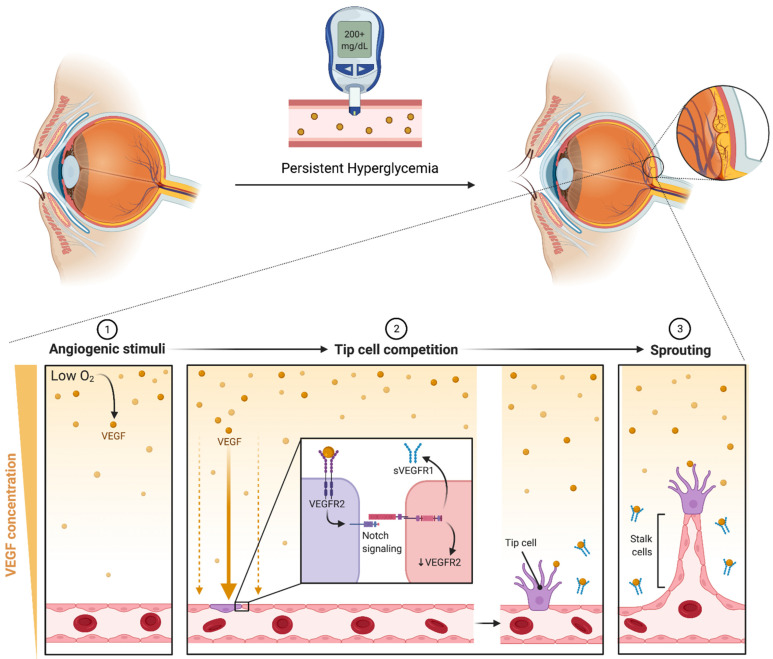
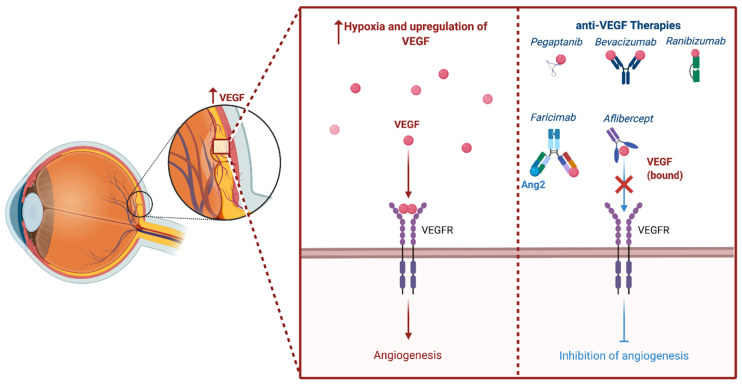
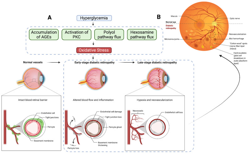
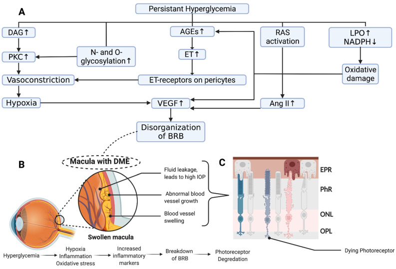
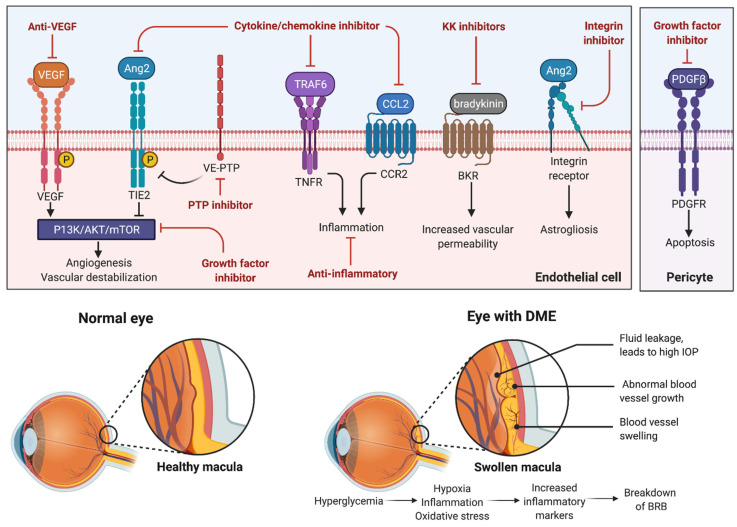
Diabetic macular edema (DME) is a major ocular complication of diabetes mellitus (DM), leading to significant visual impairment. DME's pathogenesis is multifactorial. Focal edema tends to occur when primary metabolic abnormalities lead to a persistent hyperglycemic state, causing the development of microaneurysms, often with extravascular lipoprotein in a circinate pattern around the focal leakage. On the other hand, diffusion edema is due to a generalized breakdown of the inner blood-retinal barrier, leading to profuse early leakage from the entire capillary bed of the posterior pole with the subsequent extravasation of fluid into the extracellular space. The pathogenesis of DME occurs through the interaction of multiple molecular mediators, including the overexpression of several growth factors, including vascular endothelial growth factor (VEGF), insulin-like growth factor-1, angiopoietin-1, and -2, stromal-derived factor-1, fibroblast growth factor-2, and tumor necrosis factor. Synergistically, these growth factors mediate angiogenesis, protease production, endothelial cell proliferation, and migration. Treatment for DME generally involves primary management of DM, laser photocoagulation, and pharmacotherapeutics targeting mediators, namely, the anti-VEGF pathway. The emergence of anti-VEGF therapies has resulted in significant clinical improvements compared to laser therapy alone. However, multiple factors influencing the visual outcome after anti-VEGF treatment and the presence of anti-VEGF non-responders have necessitated the development of new pharmacotherapies. In this review, we explore the pathophysiology of DME and current management strategies. In addition, we provide a comprehensive analysis of emerging therapeutic approaches to the treatment of DME.

Keywords: VEGF; diabetic macular edema; intravitreal injection; laser photocoagulation; novel pharmacotherapy; therapeutics.


Conflict of interest statement

The authors declare no conflict of interest.

Figures

Figure 3
Figure 3
Persistent hyperglycemia causes retinal vascular structural changes that lead to hypoperfusion and decreased oxygen content. Low oxygen levels induce VEGF production in tissues, which binds to VEGF receptor-2 (VEGFR2) on the surface of endothelial cells triggering differentiation into tip cells. Inhibition of neighboring tip cell formation is carried out through notch signaling, which downregulates VEGFR2 and induces expression of soluble VEGF receptor-1 (sVEGFR1). sVEGFR1 serves to downregulate angiogenic VEGF-VEGFR2 signaling, thereby preventing excessive vascularization. Neighboring cells form the body of the sprouting vessel becoming stalk cells. It has been postulated that sVEGFR-1 functions as a guide molecule in vascular sprouting through inactivating VEGF on both sides of the sprout, creating a VEGF-rich path ensuring that the stalk grows in the correct direction [78,79] (created with BioRender).
Figure 4
Figure 4
The phrase “anti-VEGF” encompasses a variety of distinct compounds that inhibit angiogenesis. The four most studied structural compounds include aptamers (i.e., pegaptanib), antibodies to VEGF (i.e., bevacizumab), antibody fragments against VEGF (i.e., ranibizumab), fusion proteins (i.e., aflibercept), and heteroantibodies (i.e., Faricimab) (created with BioRender).
Figure 1
Figure 1
(A) Chronic hyperglycemia in diabetes leads to the upregulation of pathways, activation of enzymes, and accumulation advanced glycosylated end products. Each of these pathways can incite oxidative stress and set off the pathogenesis of diabetic retinopathy, which is characterized by loss of vascular endothelial cells, tight junctions, and pericytes, with basement membrane thickening, ultimately leading to hypoxia and neovascularization. (B) This can be observed in the retina as dot blot hemorrhages, cotton wool spots, and hard exudates in the outer plexiform layer (created with BioRender).
Figure 2
Figure 2
(A) Schematic overview of potential mechanisms leading to diabetic macular edema. Hyperglycemia-induced metabolic stress leads to a complex interaction leading to vascular damage to and compromise of the blood–retinal barrier (BRB) (Modified from [29]). (B) Disorganization of the BRB and hypoperfusion causes fluid extravasation, neovascularization, and subsequent edema. (C) Chronic swelling of the macula leads to damage to the neural retina and loss of photoreceptors (created with BioRender).
Figure 5
Figure 5
This figure provides an overview of innovative therapeutic approaches that target distinct signaling pathways implicated in the pathophysiology of DME.

References

    1. Kumar V., Abbas A.K., Aster J.C., Perkins J.A. Robbins Basic Pathology. 10th ed. Elsevier; Philadelphia, PA, USA: 2018. p. xiv.935p
    1. GBD 2019 Blindness and Vision Impairment Collaborators on behalf of the Vision Loss Expert Group of the Global Burden of Disease Study Causes of blindness and vision impairment in 2020 and trends over 30 years, and prevalence of avoidable blindness in relation to VISION 2020: The Right to Sight: An analysis for the Global Burden of Disease Study. Lancet Glob. Health. 2021;9:e144–e160. doi: 10.1016/S2214-109X(20)30489-7. - DOI - PMC - PubMed
    1. Liu R., Ma B., Gao Y., Ma B., Liu Y., Qi H. Tear Inflammatory Cytokines Analysis and Clinical Correlations in Diabetes and Nondiabetes With Dry Eye. Am. J. Ophthalmol. 2019;200:10–15. doi: 10.1016/j.ajo.2018.12.001. - DOI - PubMed
    1. Wiemer N.G., Dubbelman M., Kostense P.J., Ringens P.J., Polak B.C. The influence of chronic diabetes mellitus on the thickness and the shape of the anterior and posterior surface of the cornea. Cornea. 2007;26:1165–1170. doi: 10.1097/ICO.0b013e31814fa82f. - DOI - PubMed
    1. Spaide R.F. Measurable Aspects of the Retinal Neurovascular Unit in Diabetes, Glaucoma, and Controls. Am. J. Ophthalmol. 2019;207:395–409. doi: 10.1016/j.ajo.2019.04.035. - DOI - PubMed

MeSH terms

Substances