Review
. 2022 Jun 18;11(12):1963.
doi: 10.3390/cells11121963.

Dysregulated Ca2+ Homeostasis as a Central Theme in Neurodegeneration: Lessons from Alzheimer's Disease and Wolfram Syndrome

Affiliations
Review

Dysregulated Ca2+ Homeostasis as a Central Theme in Neurodegeneration: Lessons from Alzheimer's Disease and Wolfram Syndrome

Manon Callens et al. Cells. .

Abstract

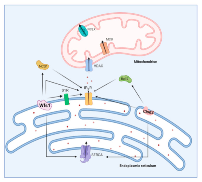
Calcium ions (Ca2+) operate as important messengers in the cell, indispensable for signaling the underlying numerous cellular processes in all of the cell types in the human body. In neurons, Ca2+ signaling is crucial for regulating synaptic transmission and for the processes of learning and memory formation. Hence, the dysregulation of intracellular Ca2+ homeostasis results in a broad range of disorders, including cancer and neurodegeneration. A major source for intracellular Ca2+ is the endoplasmic reticulum (ER), which has close contacts with other organelles, including mitochondria. In this review, we focus on the emerging role of Ca2+ signaling at the ER-mitochondrial interface in two different neurodegenerative diseases, namely Alzheimer's disease and Wolfram syndrome. Both of these diseases share some common hallmarks in the early stages, including alterations in the ER and mitochondrial Ca2+ handling, mitochondrial dysfunction and increased Reactive oxygen species (ROS) production. This indicates that similar mechanisms may underly these two disease pathologies and suggests that both research topics might benefit from complementary research.

Keywords: Alzheimer’s disease; Wolfram syndrome; calcium; mitochondria; mitochondria-associated ER membranes (MAMs); neurodegeneration.


Conflict of interest statement

The authors declare no conflict of interest.

Figures

Figure 1
Figure 1
(A) Schematic representation of the Ca2+-signaling toolkit. Ca2+ can enter the cell via store-operated, STIM-gated Orai channels, but also through voltage-gated Ca2+ channels (VOC), arachidonic acid-regulated Ca2+ channels (ARC), transient receptor potential channels (TRP) and ligand-gated ion channels (LGIC). The plasma membrane Ca2+ ATPase (PMCA) and the plasmalemmal Na+/Ca2+ exchanger (NCX) are responsible for Ca2+ extrusion into the extracellular space. Ca2+ is mainly stored in intracellular Ca2+ stores, such as the endoplasmic reticulum, where it is buffered by calreticulin (CRT) and calnexin (CNX). Mobilization of Ca2+ from the ER into the cytosol occurs via two main channels, namely the inositol 1,4,5-trisphosphate receptor (IP3R) and the ryanodine receptor (RyR). Multiple ER-Ca2+ leak channels exist including presenilin (PSEN), Bak inhibitor-1 (BI-1), TRP melastin 8 (TRPM8) and Sec61 translocon. ER Ca2+ depletion is detected by luminal ER Ca2+ sensors STIM1/STIM2, which subsequently activate plasmalemmal Orai to cause a Ca2+ influx across the plasma membrane. (Re)filling of the ER is mediated by the sarco-endoplasmic reticulum ATPase (SERCA). In the mitochondria, Ca2+ uptake is mediated by the voltage-gated anion channel (VDAC) on the outer mitochondrial membrane and the mitochondrial calcium uniporter (MCU) complex on the inner mitochondrial membrane. The main mitochondrial Ca2+ efflux transporter is the Na+/Ca2+ exchanger (NCLX). Lysosomal Ca2+ release is mediated by transient receptor potential mucolipin (TRPML) and two-pore channel 2 (TPC2). This latter is regulated by nicotinic acid adenine dinucleotide phosphate (NAADP). Cytosolic Ca2+ is buffered by several Ca2+-binding proteins including calmodulin (CaM), calbindin D-28 (CB-D28K), calretinin (CR) and parvalbumin (PV); (BD): Schematic overview of relevant examples of functional correlates of Ca2+ signaling in specific tissue/cell types. CICR, Ca2+-induced Ca2+ release; SR, sarcoplasmic reticulum; ER, endoplasmic reticulum; AMPA, α-amino-3-hydroxy-5-methyl-4-isoxazolepropionic acid; NMDA, N-methyl-D-aspartate; LTP, long term potentiation; TRPM5, transient receptor potential cation channel subfamily M member 5.
Figure 2
Figure 2
Schematic overview of changes in ER Ca2+ handling occurring in Alzheimer’s disease. Mutations in presenilin (PSEN) were shown to increase the expression of the RyR and to cause an enhancement of IP3R-mediated Ca2+ release. Additionally, the RyR is responsible for some of the IP3R-mediated Ca2+ release through CICR. PSENs were also reported to form ER Ca2+ leak channels, a function that is abolished by PSEN mutations and which causes ER Ca2+ overload. PSEN mutations also display an inhibitory effect on store-operated Ca2+ entry, which may be involved in the generation of toxic Aβ deposits. Finally, PSENs interact with SERCA which also impacts the Aβ formation.
Figure 3
Figure 3
Schematic overview of the feed forward interplay between Ca2+ signaling and Aβ. Mutations in presenilin (PSEN) cause dysregulations in Ca2+ homeostasis, which then causes altered processing of APP and the formation of Aβ. In turn, this further impacts Ca2+ homeostasis.
Figure 4
Figure 4
Representation of the mitochondrial changes reported in Alzheimer’s disease. Aβ has been shown to induce mitochondrial Ca2+ overload via different mechanisms. For instance, Aβ inhibits Ca2+ extrusion by blocking the Na+/Ca2+ exchanger (NCLX). On the other hand, Aβ promotes Ca2+ uptake via interaction with mitochondrial Ca2+ uniporter (MCU). Aβ deposits are also detected inside the mitochondria where they further contribute to dysregulated Ca2+ homeostasis. The uptake of Aβ in the mitochondria is reportedly regulated by translocase of the outer membrane (TOM) machinery. The mitochondrial Ca2+ overload causes increased production of ROS and further leads to mitochondrial dysfunction.
Figure 5
Figure 5
Overview of the role of two Wolfram syndrome proteins on ER Ca2+ handling. Wolframin (Wfs1) stimulates IP3R-mediated Ca2+ release both directly and via Neuronal Ca2+ Sensor 1 (NCS1). On the other hand, Cisd2 also interacts with the IP3R in a direct and indirect manner. Cisd2 modulates B-cell lymphoma 2 (Bcl-2), which is a known inhibitor of the IP3R. Both Wfs1 and Cisd2 were shown to interact with sarco-endoplasmic reticulum ATPase (SERCA).
Figure 6
Figure 6
Schematic overview indicating potential therapeutic strategies to target both Alzheimer’s disease and Wolfram syndrome. Possible therapeutic targets are indicated by a red asterisk. S1R, sigma 1 receptor; Bcl-2, B-cell lymphoma 2; IP3R, inositol trisphosphate receptor; RyR, ryanodine receptor; SERCA, sarco/endoplasmic reticulum ATPase; VDAC, voltage-dependent anion channel; Wfs1, wolframin.

Similar articles

Cited by

References

    1. Berridge M.J., Lipp P., Bootman M.D. The versatility and universality of calcium signalling. Nat. Rev. Mol. Cell Biol. 2000;1:11–21. doi: 10.1038/35036035. - DOI - PubMed
    1. Bootman M.D., Bultynck G. Fundamentals of Cellular Calcium Signaling: A Primer. Cold Spring Harb. Perspect. Biol. 2020;12:a038802. doi: 10.1101/cshperspect.a038802. - DOI - PMC - PubMed
    1. Rizzuto R., De Stefani D., Raffaello A., Mammucari C. Mitochondria as sensors and regulators of calcium signalling. Nat. Rev. Mol. Cell Biol. 2012;13:566–578. doi: 10.1038/nrm3412. - DOI - PubMed
    1. Parys J.B., Guse A.H. Full focus on calcium. Sci. Signal. 2019;12:eaaz0961. doi: 10.1126/scisignal.aaz0961. - DOI - PubMed
    1. Brini M., Calì T., Ottolini D., Carafoli E. Neuronal calcium signaling: Function and dysfunction. Cell. Mol. Life Sci. 2014;71:2787–2814. doi: 10.1007/s00018-013-1550-7. - DOI - PMC - PubMed

Publication types