. 2022 Jun 19;11(12):1968.
doi: 10.3390/cells11121968.

When Electrospun Fiber Support Matters: In Vitro Ovine Long-Term Folliculogenesis on Poly (Epsilon Caprolactone) (PCL)-Patterned Fibers

Affiliations

When Electrospun Fiber Support Matters: In Vitro Ovine Long-Term Folliculogenesis on Poly (Epsilon Caprolactone) (PCL)-Patterned Fibers

Chiara Di Berardino et al. Cells. .

Abstract

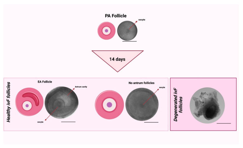
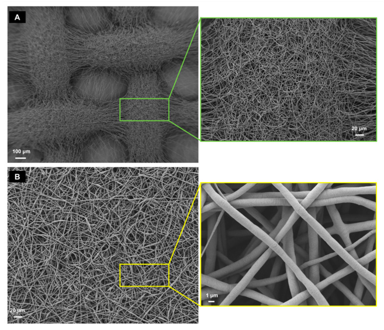
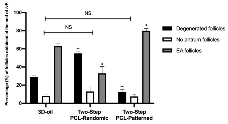
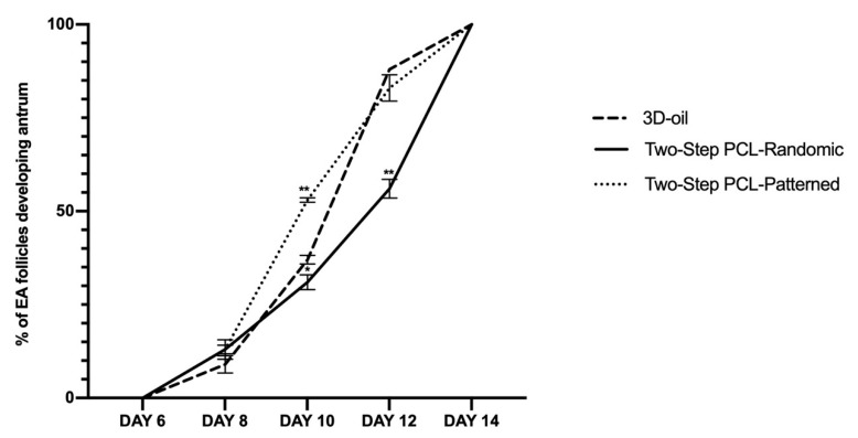
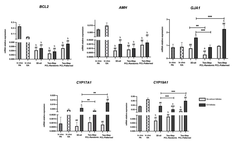
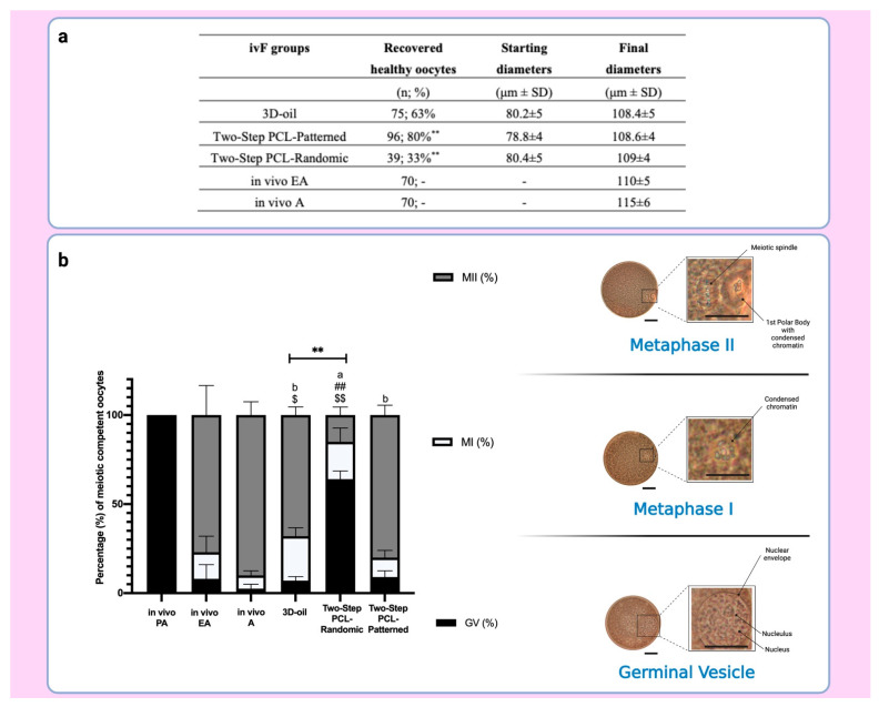
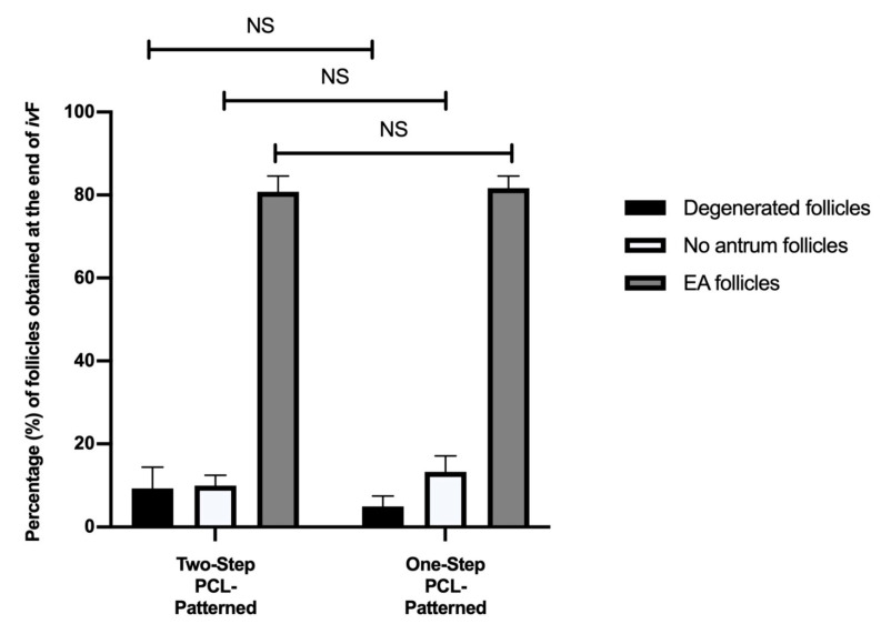
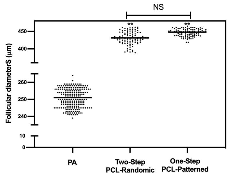
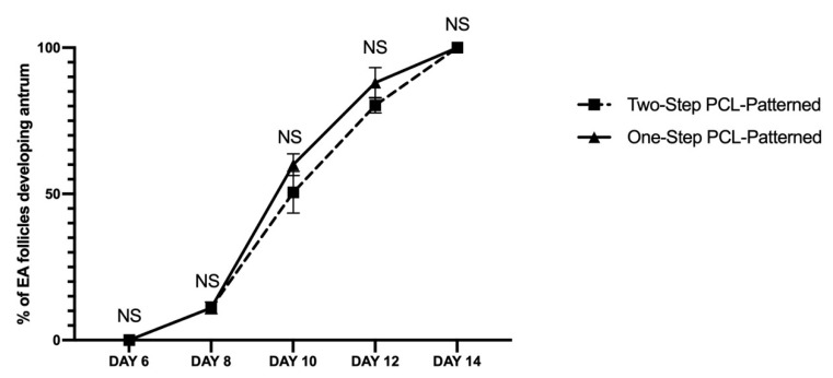
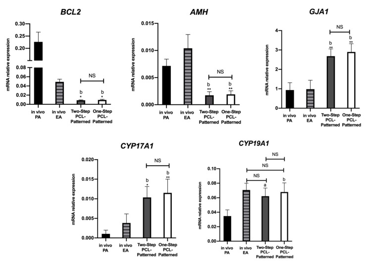
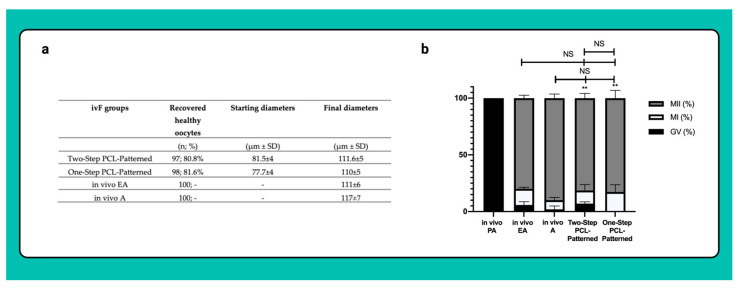
Current assisted reproduction technologies (ART) are insufficient to cover the slice of the population needing to restore fertility, as well as to amplify the reproductive performance of domestic animals or endangered species. The design of dedicated reproductive scaffolds has opened the possibility to better recapitulate the reproductive 3D ovarian environment, thus potentially innovating in vitro folliculogenesis (ivF) techniques. To this aim, the present research has been designed to compare ovine preantral follicles in vitro culture on poly(epsilon-caprolactone) (PCL)-based electrospun scaffolds designed with different topology (Random vs. Patterned fibers) with a previously validated system. The ivF performances were assessed after 14 days under 3D-oil, Two-Step (7 days in 3D-oil and on scaffold), or One-Step PCL protocols (14 days on PCL-scaffold) by assessing morphological and functional outcomes. The results show that Two- and One-Step PCL ivF protocols, when performed on patterned scaffolds, were both able to support follicle growth, antrum formation, and the upregulation of follicle marker genes leading to a greater oocyte meiotic competence than in the 3D-oil system. In conclusion, the One-Step approach could be proposed as a practical and valid strategy to support a synergic follicle-oocyte in vitro development, providing an innovative tool to enhance the availability of matured gametes on an individual basis for ART purposes.

Keywords: PCL-electrospun scaffold; artificial-ovary; in vitro follicle culture; oocyte meiotic competence; patterned fibers; preantral follicle growth; random fibers; sheep.


Conflict of interest statement

The authors declare no conflict of interest.

Figures

Figure 1
Figure 1
ivF culture protocols carried out on single follicle. Two-Step culture protocols with PCL-Randomic and PCL-Patterned fibrous scaffold were compared to the previously validated 3D-oil protocol. Secondly, Two-Step and One-Step PCL-Patterned approaches performances were compared. Specifically: (A) in 3D-oil, single PA follicles were placed in a 25 μL drop of culture medium under a 75 μL drop of pre-equilibrated mineral oil (density 5 0.84 g/mL) and then overlaid by 25 μL oil in a 96-well dish with V-shaped wells; (B) in Two-Step, single PA follicles were placed in a 25 μL drop of culture medium under a 75 μL drop of pre-equilibrated mineral oil (density 5 0.84 g/mL) and then overlaid by 25 μL oil in a 96-well dish with V-shaped wells before antrum formation (day 6 of ivF culture) to then move the growing single follicle on trans-well culture system with the holder filled with PCL-Randomic or Patterned electrospun scaffolds (PCL-Randomic vs. PCL-Patterned); (C) in One-Step, single PA follicles were in vitro cultured on the trans-well culture system with one of the electrospun PCL-scaffold identify during the (B) step for 14 days. Three independent biological replicates were performed, and for each protocol a total of 120, 125, 122, and 120 follicles were considered (respectively for 3D-oil, Two-Step PCL-Randomic, Two-Step PCL-Patterned, One-Step PCL-Patterned), and compared the ivF performances by assessing follicular (follicles in vitro development and granulosa cells’ gene expression) and germinal (acquisition of meiotic competence using IVM) compartments. Created with BioRender.com.
Figure 2
Figure 2
Comparison between healthy follicles after ivF cultures (no antrum and EA follicles) vs. unhealthy follicles after ivF cultures (degenerated follicles). Scale bar: 200 μm. Created with BioRender.com.
Figure 3
Figure 3
SEM analysis of the PCL-Patterned (A) and PCL-Randomic (B) scaffolds. PCL-Patterned scaffold is reported in A and its zoomed view is shown in a green frame. The scale bars are 100 μm (magnification 100×) and 20 μm (magnification 500×), respectively. The PCL-Randomic is reported in the bottom (B) (magnification 500×) and a higher magnification (10 k× image is shown in a yellow frame. For those images the scale bars are 10 μm and 1 μm, respectively.
Figure 4
Figure 4
Percentage (%) of degenerated, no antrum and EA follicles per groups at the end of ivF culture. A total of 120, 125, and 122 follicles were cultured in three independent experiments during which 3D-oil, Two-Step PCL-Randomic and Two-Step PCL-Patterned electrospun scaffolds were simultaneal compared (3D-oil: 40, 40 and 40 PA follicles per experiments; Two-Step PCL-Randomic: 41, 42 and 42 follicles PA follicles per experiments; Two-Step PCL-Patterned: 41, 40, and 41 PA follicles per experiments). For each ivF group (3D-oil, PCL-Randomic and PCL-Patterned electrospun scaffolds) 75, 39 and 96 EA follicles as well as 24, 44, and 10 degenerated follicles were collected at the end of in vitro culture, respectively. a,b: statistically significant vs. EA follicles of the 3D-oil group (p < 0.05 and p < 0.01, respectively); **: statistically significant vs. degenerated follicles of the 3D-oil group (p < 0.01). NS: not statistically significant.
Figure 5
Figure 5
EA follicular mean final diameters: overview for each experimental group. A total of 120, 125, and 122 follicles were cultured in three independent experiments during which 3D-oil, Two-Step PCL-Randomic, and Two-Step PCL-Patterned electrospun scaffolds were simultaneal compared (3D-oil: 40, 40, and 40 PA follicles per experiments; Two-Step PCL-Randomic: 41, 42, and 42 follicles PA follicles per experiments; Two-Step PCL-Patterned: 41, 40, and 41 PA follicles per experiments). A total of 367 follicles (showed in the graph as black dots) were measured to determine the starting mean diameters (PA). Then, 75, 39, and 96 EA follicles, respectively for 3D-oil, Two-Step PCL-Randomic, and Two-Step PCL-Patterned were measured at the end of ivF culture to determine the final mean diameters. **: statistically significant vs. PA group (p < 0.01). NS: not statistically significant.
Figure 6
Figure 6
Development trend of the follicular antrum over time according to follicular treatment groups. A total of 120, 125, and 122 follicles were cultured in three independent experiments during which 3D-oil, Two-Step PCL-Randomic, and Two-Step PCL-Patterned electrospun scaffolds were simultaneal compared (3D-oil: 40, 40 and 40 PA follicles per experiments; Two-Step PCL-Randomic: 41, 42, and 42 follicles PA follicles per experiments; Two-Step PCL-Patterned: 41, 40, and 41 PA follicles per experiments). Specifically, only follicles that differentiated the follicular antrum during ivF cultures, were tested (75, 39, and 96 EA follicles, respectively, for 3D-oil, Two-Step PCL-Randomic, and Two-Step PCL-Patterned electrospun scaffolds). *, **: statistically significant vs. 3D-oil group (p < 0.05 and p < 0.01, respectively). Data in which subscripts are not indicated, were considered not statistically significant.
Figure 7
Figure 7
Representative expression of steroidogenic and somatic-specific genes. Twelve follicular walls per group were processed for gene expression analysis. Data were expressed as mean ± SD. **, ***: statistically significant vs. 3D-oil group (p < 0.05, p < 0.01 and p < 0.0001, respectively). #, ##, ###: statistically significant vs. in vivo EA follicles (p < 0.05, p < 0.01 and p < 0.0001, respectively). a,b: statistically significant vs. in vivo PA follicles (p < 0.05 and p < 0.01, respectively). Data in which subscripts are not indicated were considered not statistically significant.
Figure 8
Figure 8
(a) Table summarizing the number of recovered healthy oocytes and their mean diameters at the end of ivF culture. **: statistically significant vs. 3D-oil group. (b) On the left: Percentage of the oocyte meiotic competence acquired in vitro. Different nuclear stages were characterized. a,b: statistically significant vs. recovered healthy oocytes from in vivo PA (p < 0.05 and p < 0.01, respectively); ##: statistically significant vs. recovered healthy oocytes from in vivo EA (p < 0.01); $, $$: statistically significant vs. recovered healthy oocytes from in vivo A (p < 0.05 and p < 0.01, respectively); **: statistically significant vs. recovered healthy oocytes from 3D-oil group (p < 0.01). Data in which subscripts are not indicated, were considered not statistically significant. On the right: Images of oocyte nuclear stages. GV: Germinal Vesicle Break Down; MI: Metaphase 1; MII: Metaphase II (Lacmoid solution staining, 40× magnification: scale bar: 30 μm; insert of the oocytes details; scale bar 30 μm). Created with BioRender.com.
Figure 9
Figure 9
Percentage (%) of degenerated, no antrum and EA follicles per groups at the end of ivF culture. A total of 120 and 120 follicles were cultured in three independent experiments during which Two-Step PCL-Patterned and One-step PCL-Patterned electrospun scaffolds were simultaneal compared (Two-Step PCL-Patterned: 40, 40, and 40 PA follicles per experiment; One-Step PCL-Patterned: 40, 40, and 40 PA follicles per experiment). NS: not statistically significant.
Figure 10
Figure 10
EA follicular mean final diameters: overview for each experimental group. A total of 120 and 120 follicles were cultured in three independent experiments during which Two-Step PCL-Patterned and One-step PCL-Patterned electrospun scaffolds were simultaneal compared (Two-Step PCL-Patterned: 40, 40, and 40 PA follicles per experiment; One-Step PCL-Patterned: 40, 40, and 40 PA follicles per experiment). **: statistically significant vs. PA follicles group (p < 0.01). NS: not statistically significant.
Figure 11
Figure 11
Development trend of the follicular antrum over time according to follicular treatment groups. A total of 120 and 120 follicles were cultured in three independent experiments during which Two-Step PCL-Patterned and One-step PCL-Patterned electrospun scaffolds were simultaneal compared (Two-Step PCL-Patterned: 40, 40, and 40 PA follicles per experiment; One-Step PCL-Patterned: 40, 40, and 40 PA follicles per experiment). Specifically, only follicles that differentiated the follicular antrum during ivF cultures, were tested (97 and 98 EA follicles, respectively, for Two-Step PCL-Patterned and One-Step PCL-Patterned). NS: not statistically significant.
Figure 12
Figure 12
Representative expression of steroidogenic and somatic-specific genes. Twelve follicular walls per group were processed for gene expression analysis. Data were expressed as mean ± SD. a,b: statistically significant vs. in vivo PA follicles group (p < 0.05 and p < 0.01, respectively). *, **: statistically significant vs. in vivo EA follicles group (p < 0.05 and p < 0.01, respectively). NS: not statistically significant.
Figure 13
Figure 13
(a) Table summarizing the number of recovered healthy oocytes and their mean diameters at the end of ivF culture. A total of 120 and 120 follicles were cultured in three independent experiments during which Two-Step PCL-Patterned and One-step PCL-Patterned electrospun scaffolds were simultaneal compared (Two-Step PCL-Patterned: 40, 40, and 40 PA follicles per experiment; One-Step PCL-Patterned: 40, 40, and 40 PA follicles per experiment). For each ivF group (Two-Step PCL-Patterned and One-Step PCL-Patterned), 97 and 98 healthy oocytes were recovered and analyzed at the end of the in vitro culture to test the oocyte performance. Only the oocytes retrieved from follicles that differentiated the follicular antrum, were tested; data are expressed as mean ± SD. (b) Percentage of the oocyte meiotic competence acquired in vitro. Different nuclear stages were characterized. **: statistically significant vs. in vivo PA follicles group (p < 0.05). NS: not statistically significant. Data in which subscripts are not indicated, were considered not significant. GV: Germinal Vesicle; MI: Metaphase I; MII: Metaphase II.

Similar articles

Cited by

References

    1. Dolmans M.M., Amorim C.A. Construction and Use of Artificial Ovaries. Reproduction. 2019;158:F15–F25. doi: 10.1530/REP-18-0536. - DOI - PubMed
    1. Gougeon A. Regulation of Ovarian Follicular Development in Primates: Facts and Hypotheses. Endocr. Rev. 1996;17:121–155. doi: 10.1210/edrv-17-2-121. - DOI - PubMed
    1. Scaramuzzi R.J., Baird D.T., Campbell B.K., Driancourt M.A., Dupont J., Fortune J.E., Gilchrist R.B., Martin G.B., McNatty K.P., McNeilly A.S., et al. Regulation of Folliculogenesis and the Determination of Ovulation Rate in Ruminants. Reprod. Fertil. Dev. 2011;23:444–467. doi: 10.1071/RD09161. - DOI - PubMed
    1. Adamson G.D., de Mouzon J., Chambers G.M., Zegers-Hochschild F., Mansour R., Ishihara O., Banker M., Dyer S. International Committee for Monitoring Assisted Reproductive Technology: World Report on Assisted Reproductive Technology, 2011. Fertil. Steril. 2018;110:1067–1080. doi: 10.1016/j.fertnstert.2018.06.039. - DOI - PubMed
    1. Ori S. Global Trends in Reproductive Policy and Practice. 8th ed. Volume 4. IFFS; Royal, NJ, USA: 2019. International Federation of Fertility Societies’ Surveillance International Federation of Fertility Societies’ Surveillance (IFFS) - DOI

Publication types

LinkOut - more resources