

Imaging Features of Post Main Hepatectomy Complications: The Radiologist Challenging
- PMID: 35741133
- PMCID: PMC9221607
- DOI: 10.3390/diagnostics12061323
Imaging Features of Post Main Hepatectomy Complications: The Radiologist Challenging
Abstract
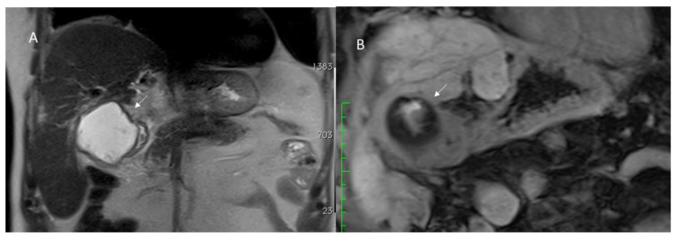
In the recent years, the number of liver resections has seen an impressive growth. Usually, hepatic resections remain the treatment of various liver diseases, such as malignant tumors, benign tumors, hydatid disease, and abscesses. Despite technical advancements and tremendous experience in the field of liver resection of specialized centers, there are moderately high rates of postoperative morbidity and mortality, especially in high-risk and older patient populations. Although ultrasonography is usually the first-line imaging examination for postoperative complications, Computed Tomography (CT) is the imaging tool of choice in emergency settings due to its capability to assess the whole body in a few seconds and detect all possible complications. Magnetic resonance cholangiopancreatography (MRCP) is the imaging modality of choice for delineating early postoperative bile duct injuries and ischemic cholangitis that may arise in the late postoperative phase. Moreover, both MDCT and MRCP can precisely detect tumor recurrence. Consequently, radiologists should have knowledge of these surgical procedures for better comprehension of postoperative changes and recognition of the radiological features of various postoperative complications.
Keywords: hepatectomy; postoperative complications; radiologists.
Conflict of interest statement
The authors have no conflict of interest to disclose. The authors confirm that the article is not under consideration for publication elsewhere.
Figures


References
-
- Granata V., Grassi R., Fusco R., Setola S.V., Belli A., Ottaiano A., Nasti G., La Porta M., Danti G., Cappabianca S., et al. Intrahepatic cholangiocarcinoma and its differential diagnosis at MRI: How radiologist should assess MR features. Radiol. Med. 2021;126:1584–1600. doi: 10.1007/s11547-021-01428-7. - DOI - PubMed
Publication types
LinkOut - more resources
Full Text Sources
