Review
. 2022 Jun 3;12(6):1386.
doi: 10.3390/diagnostics12061386.

Neurogenic Appendicitis: A Reappraisal of the Clinicopathological Features and Pathogenesis

Affiliations
Review

Neurogenic Appendicitis: A Reappraisal of the Clinicopathological Features and Pathogenesis

Mahmoud Rezk Abdelwahed Hussein et al. Diagnostics (Basel). .

Abstract

In 1921; Masson and Maresch first coined the term "neurogenic appendicitis (NA)" to describe "neuroma-like" lesions in the appendix. To date, our knowledge about NA is limited; therefore, we conducted a comprehensive analysis of the literature (1921 to 2020) to examine the clinicopathological features of NA. We also addressed the pathophysiology of acute abdominal pain and fibrosis in this entity. We performed a meta-analysis study by searching the PubMed database, using several keywords, such as: "appendix," "neurogenic," "obliterative," "neuroma," "fibrous obliteration," "appendicopathy," and "appendicitis." Our study revealed that patients with NA usually present clinically with features of acute appendicitis, bud2t they have grossly unremarkable appendices. Histologically, the central appendiceal neuroma was the most common histological variant of NA, followed by the submucosal and intramucosal variants. To conclude, NA represents a form of neuroinflammation. The possibility of NA should be considered in patients with clinical features of acute appendicitis who intraoperatively show a grossly unremarkable appendix. Neuroinflammation and neuropeptides play roles in the development of pain and fibrosis in NA.

Keywords: appendix; neurogenic; neuroinflammation.


Conflict of interest statement

The authors declare no conflict of interest.

Figures
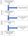
Figure 1
Figure 1
Flow chart of literature search and study selection for cases of neurogenic appendicitis.
Figure 2
Figure 2
The histological features of the central variant of neurogenic appendicitis (neuroma of the appendix). Different magnifications of the appendix reveal obliteration of the lumen, effacement of the appendiceal micro-architecture, with loss of the mucosal glands and the lymphoid follicles (AF). There is an obliteration of the lumen by residual reactive lymphoid aggregates (long arrow), and varying proportions of nerve tangles, fibrous tissue, collagen, fat, and chronic inflammatory cells (AC). The presence of residual lymphoid cells is indicative of repeated attacks of minimal asymptomatic mucosal inflammation. Hypertrophied nerve bundles (short arrow) and ganglion cells (D: arrowhead) are noted. The neuronal component is composed of mitotically inactive cells, with spindly nuclei and pale cytoplasm (Schwann-like cells) obliterating the lumen and expanding the lamina propria (E,F). The hypertrophied myenteric plexuses (G) and nerve fibers (H,I) are stained with antibodies against S100 (H: red star). Synaptophysin immunostain decorates the prominent nerve plexuses, fibers, and ganglion cells (I). (Original magnifications, A: ×20, B: ×40, C: ×100, D: ×200, E: ×200, F: ×400, G: ×20, H: ×200, and I: ×400).
Figure 3
Figure 3
The histological features of the central and submucosal variants of neurogenic appendicitis. (AC): Sections from the appendix showing the formation of a small central neuroma (a central variant of neurogenic appendicitis). The histological features include obliteration of the lumen by the admixture of fibrous and neural elements (A: star) with extensive submucosal aggregates of mature adipose tissue (B) and hypertrophied nerve bundles (C: arrowhead). (D): Grossly, the appendix measures 3.5 cm in length ×1.0 cm in circumference with attached mesoappendix (2× 0.5 × 0.5 cm). The serosal surface is smooth and glistening. There are no perforations of the appendiceal wall or purulent materials. The mesoappendix has a yellow appearance, without areas of hemorrhage or necrosis. (EH) On microscopy, sections reveal features of the submucosal variant of neurogenic appendicitis including extensive loss of the mucosal glands, crypts, and lymphoid follicles, associated with the expansion of the submucosa by the admixture of fibrous and neural tissues (blue short arrow) with hypertrophied nerve bundles (neurogenous hyperplasia, long blue arrow) that are reactive for S100 immunostain (I). The presence of lymphomononuclear cells is indicative of repeated attacks of minimal subclinical mucosal inflammation (Original magnifications, A: ×20, B: ×200, C: ×200, E: ×20, F: ×40, G: ×200, H: ×400, and I: ×200).
Figure 4
Figure 4
The histological features of the intramucosal variant of neurogenic appendicitis. (AD): Sections from the appendix show intramucosal lesion with the patent lumen, destruction of the mucosal glands, and crypts expansion of the submucosa by residual lymphoid infiltrate, fibrous and neural tissues, and hypertrophied nerve bundles that extend into the mucosa. There are lobules of mature fat cells in the submucosa. The presence of lymphomononuclear cells is indicative of repeated attacks of minimal asymptomatic mucosal inflammation. No acute inflammatory cells are seen. There is no evidence of periappendicitis. (E,F): the hypertrophied never bundles and ganglion cells are consistently positive for synaptophysin. (Original magnifications, A: ×20, B: ×40, C: ×100, D: ×200, E: ×100, and F: ×400).

References

    1. Buschard K., Kjaeldgaard A. Investigation and analysis of the position, fixation, length and embryology of the vermiform appendix. Acta Chir. Scand. 1973;139:293–298. - PubMed
    1. Schumpelick V., Dreuw B., Ophoff K., Prescher A. Appendix and cecum. Embryology, anatomy, and surgical applications. Surg. Clin. N. Am. 2000;80:295–318. doi: 10.1016/S0039-6109(05)70407-2. - DOI - PubMed
    1. Stanley M.W., Cherwitz D., Hagen K., Snover D.C. Neuromas of the appendix. A light-microscopic, immunohistochemical and electron-microscopic study of 20 cases. Am. J. Surg. Pathol. 1986;10:801–815. doi: 10.1097/00000478-198611000-00008. - DOI - PubMed
    1. Olsen B.S., Holck S. Neurogenous hyperplasia leading to appendiceal obliteration: An immunohistochemical study of 237 cases. Histopathology. 1987;11:843–849. doi: 10.1111/j.1365-2559.1987.tb01887.x. - DOI - PubMed
    1. Franke C. Neurogenic appendicopathy: A common, nearly unknown disease picture. Evaluation of 816 appendices and review of the literature. Chirurg. 2001;72:1508–1509. doi: 10.1007/s001040170019. - DOI - PubMed

LinkOut - more resources