. 2022 Jun 6;12(6):1400.
doi: 10.3390/diagnostics12061400.

Deep-Learning-Based Thrombus Localization and Segmentation in Patients with Posterior Circulation Stroke

Affiliations

Deep-Learning-Based Thrombus Localization and Segmentation in Patients with Posterior Circulation Stroke

Riaan Zoetmulder et al. Diagnostics (Basel). .

Abstract

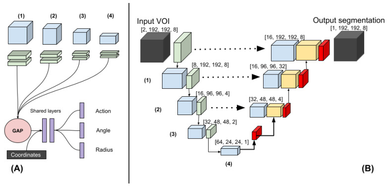
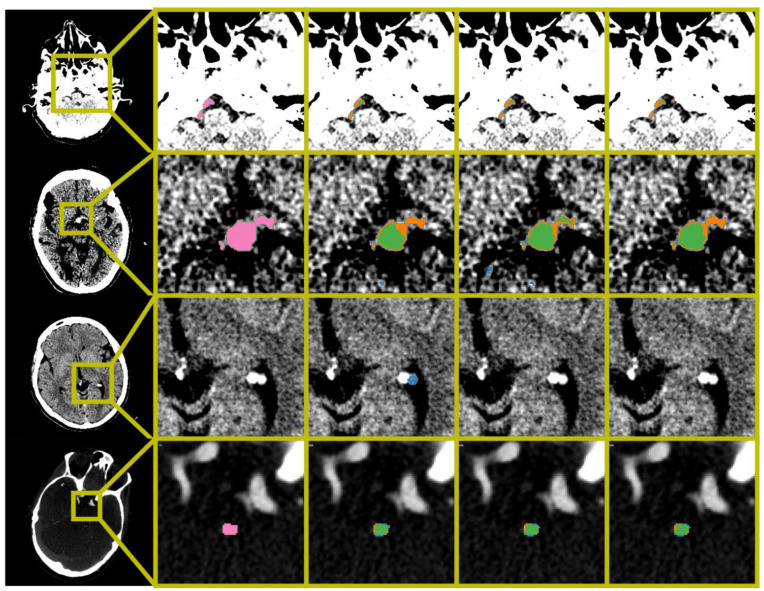
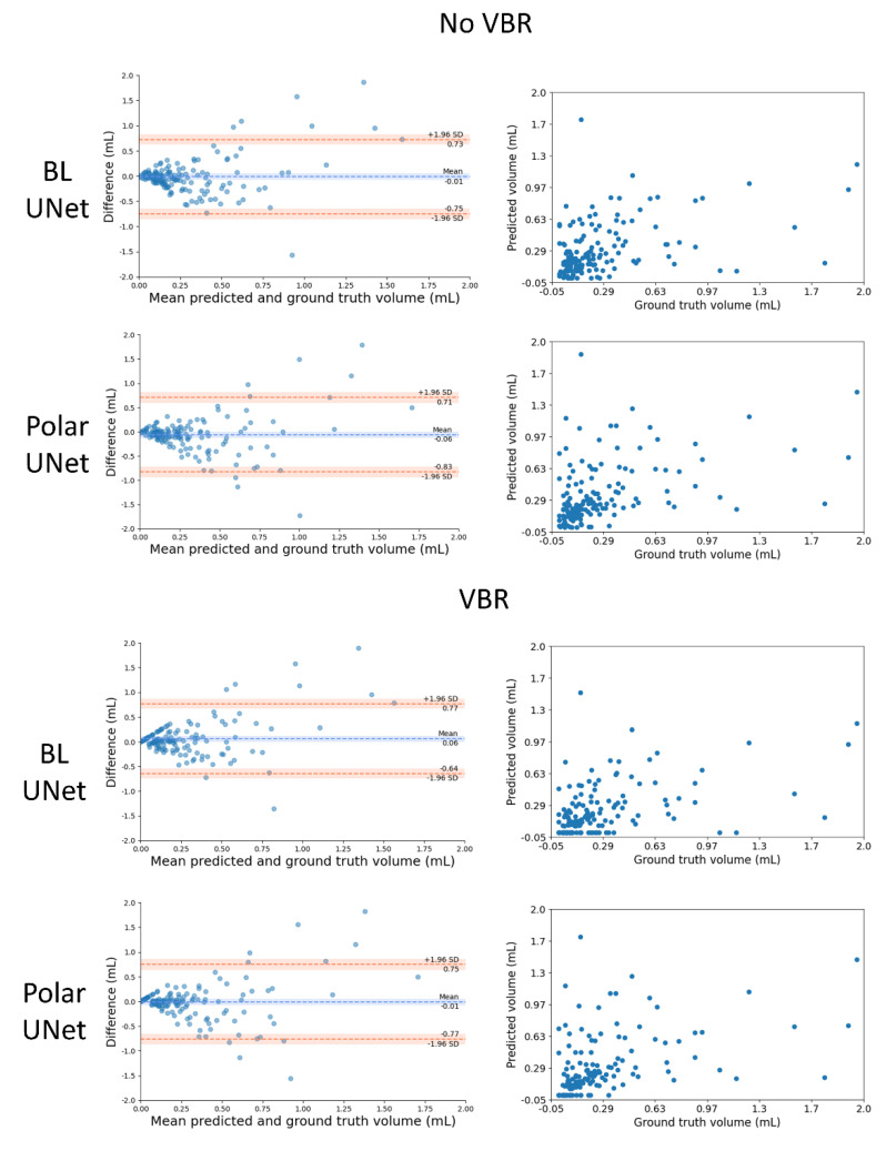
Thrombus volume in posterior circulation stroke (PCS) has been associated with outcome, through recanalization. Manual thrombus segmentation is impractical for large scale analysis of image characteristics. Hence, in this study we develop the first automatic method for thrombus localization and segmentation on CT in patients with PCS. In this multi-center retrospective study, 187 patients with PCS from the MR CLEAN Registry were included. We developed a convolutional neural network (CNN) that segments thrombi and restricts the volume-of-interest (VOI) to the brainstem (Polar-UNet). Furthermore, we reduced false positive localization by removing small-volume objects, referred to as volume-based removal (VBR). Polar-UNet is benchmarked against a CNN that does not restrict the VOI (BL-UNet). Performance metrics included the intra-class correlation coefficient (ICC) between automated and manually segmented thrombus volumes, the thrombus localization precision and recall, and the Dice coefficient. The majority of the thrombi were localized. Without VBR, Polar-UNet achieved a thrombus localization recall of 0.82, versus 0.78 achieved by BL-UNet. This high recall was accompanied by a low precision of 0.14 and 0.09. VBR improved precision to 0.65 and 0.56 for Polar-UNet and BL-UNet, respectively, with a small reduction in recall to 0.75 and 0.69. The Dice coefficient achieved by Polar-UNet was 0.44, versus 0.38 achieved by BL-UNet with VBR. Both methods achieved ICCs of 0.41 (95% CI: 0.27-0.54). Restricting the VOI to the brainstem improved the thrombus localization precision, recall, and segmentation overlap compared to the benchmark. VBR improved thrombus localization precision but lowered recall.

Keywords: CTA; NCCT; deep learning; localization; posterior stroke; segmentation; thrombus.


Conflict of interest statement

H.A. Marquering is a cofounder and shareholder of Nico.Lab. I. Išgum is a cofounder, scientific lead, and shareholder of Quantib-U. C.B.L.M. Majoie is a shareholder of Nico.Lab and has received speakers’ bureau fees from Stryker (paid to institution). D.W.J. Dippel received honoraria from Stryker (paid to institution). W.H. van Zwam received speaker fees from Stryker, NicoLab, and Cernovus, and consultation fees from Philips (all paid to institution). S.A.P. Cornelissen has no competing interests.

Figures

Figure 1
Figure 1
(A) The polar-UNet is created by attaching the following operations to the BL-UNet. The features from four levels in the down-sampling path are input to four blocks consisting of two convolutions. The output is global average-pooled and concatenated before being passed to the fully connected shared layers. Finally, individual fully connected layers are used to classify the action and regress the angle and the radius. (B) Three-dimensional Baseline UNet (BL-UNet). Three-dimensional ResNet blocks followed by a max-pooling operation (green) were used to construct the down-sampling path (left). Three-dimensional ResNet blocks followed by a transposed convolution were used to construct the up-sampling path (right). The features generated in the down- and up-sampling paths are blue and yellow, respectively. Skip connections were added between the up-sampling and down-sampling paths.
Figure 2
Figure 2
Examples of automatic segmentation results obtained by BL-UNet and Polar-UNet. From the left to right column: The original scan with a bounding box indicating the zoom location, the ground truth segmentation map, the results obtained from the BL-UNet without volume-based removal (VBR), the results obtained from the Polar-UNet without VBR, and the results obtained from the Polar-UNet with VBR. The top three rows display NCCT scans; the bottom row shows a CTA scan. The top row shows the difficulty all CNN methods have with segmenting a thrombus in the vertebral arteries. The second row from the top shows an example of small false positives removed by the VBR step. The third row from the top row shows false positives that are removed by restricting the volume-of-interest to the posterior circulation with Polar-UNet. The bottom row shows an example of a scan without a hyperdense artery sign. The segmentation maps show the ground truth (pink), true positive (green), false negative (orange) and false positive (blue). The NCCT scans were plotted using a window center level of 35, with a window width of 30. The CTA scan was plotted using a window center level of 300, with a window width of 600.
Figure 3
Figure 3
Comparison of the automated and manually segmented volume for the BL-UNet and Polar-UNet with and without volume-based removal (VBR). Left column: Bland–Altman plots of the lesion volumes. The volumes corresponding to the reference and automatic segmentations are shown on the x-axis, and the volume difference is shown on the y-axis. Right column: scatter plots comparing lesion volumes derived from the reference segmentations (y-axis) and from the automatic segmentations determined by the CNN (x-axis).

References

    1. Mattle H.P., Arnold M., Lindsberg P.J., Schonewille W.J., Schroth G. Basilar artery occlusion. Lancet Neurol. 2011;10:1002–1014. doi: 10.1016/S1474-4422(11)70229-0. - DOI - PubMed
    1. Pirson F.A.V., Boodt N., Brouwer J., Bruggeman A.A.E., den Hartog S.J., Goldhoorn R.-J.B., Langezaal L.C.M., Staals J., van Zwam W.H., van der Leij C., et al. Endovascular Treatment for Posterior Circulation Stroke in Routine Clinical Practice: Results of the Multicenter Randomized Clinical Trial of Endovascular Treatment for Acute Ischemic Stroke in the Netherlands Registry. Stroke. 2021:STROKEAHA121034786. doi: 10.1161/STROKEAHA.121.034786. - DOI - PubMed
    1. Sumer M.M., Ozdemir I., Tascilar N. Predictors of outcome after acute ischemic stroke. Acta Neurol. Scand. 2003;107:276–280. doi: 10.1034/j.1600-0404.2003.02008.x. - DOI - PubMed
    1. Lever N.M., Nyström K.V., Schindler J.L., Halliday J., Wira C., Funk M. Missed opportunities for recognition of ischemic stroke in the emergency department. J. Emerg. Nurs. 2013;39:434–439. doi: 10.1016/j.jen.2012.02.011. - DOI - PubMed
    1. Arch A.E., Weisman D.C., Coca S., Nystrom K.V., Wira C.R., Schindler J.L. Missed Ischemic Stroke Diagnosis in the Emergency Department by Emergency Medicine and Neurology Services. Stroke. 2016;47:668–673. doi: 10.1161/STROKEAHA.115.010613. - DOI - PubMed

LinkOut - more resources