Case Reports
. 2022 Jun 15;12(6):1472.
doi: 10.3390/diagnostics12061472.

Secondary (Late) Developmental Dysplasia of the Hip with Displacement: From Case Studies to a Proposition for a Modified Diagnostic Path

Affiliations
Case Reports

Secondary (Late) Developmental Dysplasia of the Hip with Displacement: From Case Studies to a Proposition for a Modified Diagnostic Path

Jacek Dygut et al. Diagnostics (Basel). .

Abstract

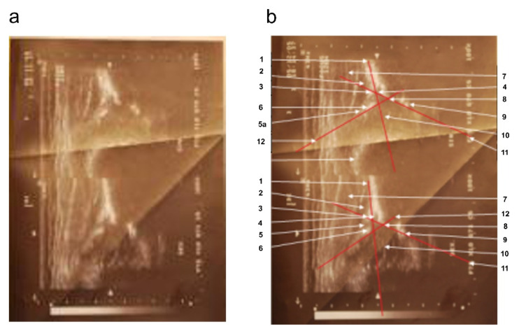
(1) Background. This paper presents a case of hip joints that were initially described as either normal or physiologically immature in four successive ultrasound examinations using the static method by Graf; however, the final treatment of the patient involved multiple hip reconstruction surgeries. (2) Case presentation. The patient was born with an Apgar score of 10 and did not exhibit neurological diseases that could deform and lead to pathological dislocation of the right hip joint. The subsequent analysis of medical data revealed that the hip luxation was due to secondary (late) developmental dysplasia of the right hip. (3) Conclusion. The article emphasizes the importance of early diagnosis and treatment standards for developmental dysplasia of the hip (DDH). The development of uniform international medical guidelines for the diagnosis, treatment, and prevention of hip dysplasia, along with the unification of DDH-related terminology, would allow for more effective management of DDH cases and reduce the cost of patient treatment.

Keywords: a case report of developmental dysplasia of the hip; diagnosis of dysplasia; treatment standards for developmental dysplasia of the hip.


Conflict of interest statement

The authors declare no conflict of interest.

Figures

Figure 1
Figure 1
Sonograms of the girl’s hip joints at 5 weeks of age (first appointment). (a) Top (left hip joint): three basic elements are visible, i.e., the labrum, the “blunt” bone edge marking the point where the bulge of the iliac bone passes into the concavity of the acetabulum, and the lower edge of the iliac bone. The alpha angle between the bone roofline and the baseline is 65°; the beta angle between the cartilaginous roofline and the baseline is 69°. Lack of ossification nuclei (physiological norm). Type Ib according to Graf. Bottom part (right hip joint): three basic elements are visible: the labrum, the “rounded” bone edge marking the point where the protrusion of the iliac bone passes into the concavity of the acetabulum, and the lower edge of the iliac bone. The alpha angle between the bone roofline and the baseline is 56°; the beta angle between the cartilage roofline and the baseline is 74°. Lack of ossification nuclei (physiological norm). Type IIa according to Graf. (b) Sonograms with overlay and description of anatomically important points. Top: 1—baseline, 2—gluteus minimus muscle, 3—tendon of the femoral rectus muscle, 4—the cartilaginous roof of the acetabulum, 5—labrum, 6—cartilaginous femoral head (no ossification nucleus), 7—cartilaginous major trochanter with the turning synovial fold of the joint capsule, 8—cartilage–bone boundary, 9—bone edge (blunt type), i.e., turning point, 10—cartilage roofline, 11—internal perichondrium of the “Y” cartilage, 12—lower ilium bone edge, 13—bone roofline, 14—soft tissues at the bottom of the acetabulum, 15—round ligament of the femoral head, 16—transverse ligament. Bottom: marked baseline, cartilage roofline, and bone roofline.
Figure 2
Figure 2
Sonograms of the girl’s hip joints at 9 weeks of age (second appointment). (a) Top part: left hip joint; alpha angle: 53°; beta angle: 74°; round bone edge. Type IIa according to Graf. Bottom: right hip joint; alpha angle: 63°; beta angle: 62°; acute bone edge type. Type Ib according to Graf. (b) Sonograms with overlay and description of anatomically important points. 1—baseline, 2—gluteus minimus muscle, 3—tendon of the femoral rectus muscle, 4—cartilage acetabular roof, 5—the disappearance of the joint capsule shadow in the region of the perichondrium defect in the adipose cushion; 5a—articular capsule, 6—acetabular labrum, 7—gluteus medium muscle, 8—bone edge (blunt type), i.e., turning point, 9—lower edge of the iliac bone, 10—cartilaginous femoral head with visualized vascular sinuses, 11—bone roofline, 12—cartilage roofline.
Figure 3
Figure 3
X-ray scan showing right hip dislocation in the girl (at 2 years of age).
Figure 4
Figure 4
Computed tomography (CT) of the pelvis—3D scan carried out at 5 years of age. The relative tilt angle of the femoral head is about 47°; the angle of internal rotation is about 25°. Knee set in internal rotation. Femoral head in anterolateral subluxation. Joint space significantly widened. Thickening of soft tissues around the hip joint; exudate cannot be excluded. Differentiation reveals postoperative changes and inflammation. The acetabulum is slightly distorted in the area of the roof and flattened with irregular contours, marginal sclerotization, and small defects. A ~4 mm calcification or ossification can be seen in the soft tissues in front of the anterior part of the acetabular roof and below the posterior edge of the roof. The femoral head is flattened. There is marked thinning of the bone structure of the femoral head and neck.
Figure 5
Figure 5
X-ray scan following varus and de-rotative osteotomy on the proximal part of the right femoral bone (at 6 years of age).

Similar articles

Cited by

References

    1. MacKenzie I.G., Wilson J.G. Problems encountered in the early diagnosis and management of congenital dislocation of the hip. J. Bone Joint. Surg. Br. 1981;63:38–42. doi: 10.1302/0301-620X.63B1.7204472. - DOI - PubMed
    1. Dunn P.M., Evans R.E., Thearle M.J., Griffiths E.D., Witherow P.J. Congenital dislocation of the hip: Early and late diagnosis. 1985 and management compared. Arch Dis. Child. 1985;60:407–414. doi: 10.1136/adc.60.5.407. - DOI - PMC - PubMed
    1. Sewell M.D., Rosendahl K., Eastwood D.M. Developmental dysplasia of the hip. BMJ. 2009;339:b4454. doi: 10.1136/bmj.b4454. - DOI - PubMed
    1. Agarwal A., Gupta N. Risk factors and diagnosis of developmental dysplasia of hip in children. J. Clin. Orthop. Trauma. 2012;3:10–14. doi: 10.1016/j.jcot.2011.11.001. - DOI - PMC - PubMed
    1. Rehabilitacja medyczna. Tom 2, wyd. II. 2012. [(accessed on 27 December 2014)]. Available online: http://www.elsevier.pl/ksiazka/rehabilitacja-medyczna-tom-2-wyd-ii.

Publication types