

Predicting Visual Acuity in Patients Treated for AMD
- PMID: 35741314
- PMCID: PMC9221868
- DOI: 10.3390/diagnostics12061504
Predicting Visual Acuity in Patients Treated for AMD
Abstract
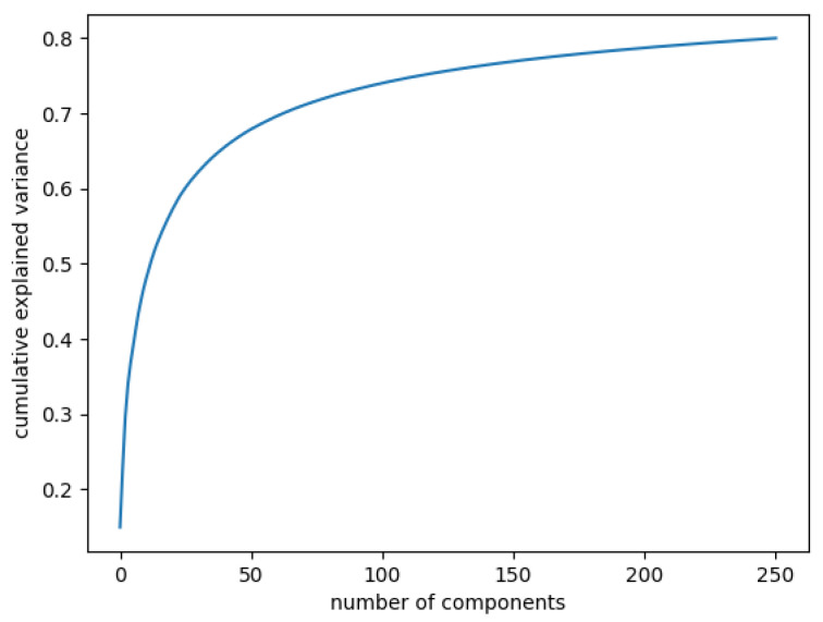
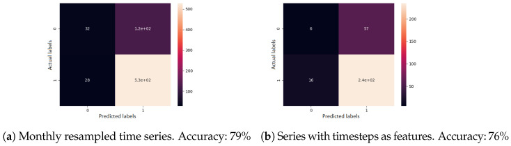
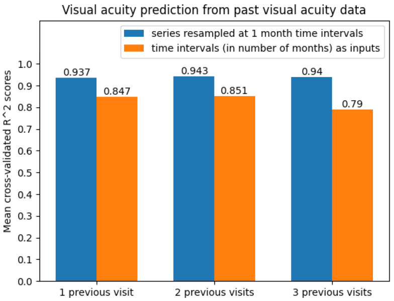
The leading diagnostic tool in modern ophthalmology, Optical Coherence Tomography (OCT), is not yet able to establish the evolution of retinal diseases. Our task is to forecast the progression of retinal diseases by means of machine learning technologies. The aim is to help the ophthalmologist to determine when early treatment is needed in order to prevent severe vision impairment or even blindness. The acquired data are made up of sequences of visits from multiple patients with age-related macular degeneration (AMD), which, if not treated at the appropriate time, may result in irreversible blindness. The dataset contains 94 patients with AMD and there are 161 eyes included with more than one medical examination. We used various techniques from machine learning (linear regression, gradient boosting, random forest and extremely randomised trees, bidirectional recurrent neural network, LSTM network, GRU network) to handle technical challenges such as how to learn from small-sized time series, how to handle different time intervals between visits, and how to learn from different numbers of visits for each patient (1-5 visits). For predicting the visual acuity, we performed several experiments with different features. First, by considering only previous measured visual acuity, the best accuracy of 0.96 was obtained based on a linear regression. Second, by considering numerical OCT features such as previous thickness and volume values in all retinal zones, the LSTM network reached the highest score (R2=0.99). Third, by considering the fundus scan images represented as embeddings obtained from the convolutional autoencoder, the accuracy was increased for all algorithms. The best forecasting results for visual acuity depend on the number of visits and features used for predictions, i.e., 0.99 for LSTM based on three visits (monthly resampled series) based on numerical OCT values, fundus images, and previous visual acuities.
Keywords: OCT; diagnosis of retinal conditions; machine learning; predicting visual acuity.
Conflict of interest statement
The authors declare they have no conflict of interest.
Figures


Similar articles
-
Use of a Neural Net to Model the Impact of Optical Coherence Tomography Abnormalities on Vision in Age-related Macular Degeneration.Am J Ophthalmol. 2018 Jan;185:94-100. doi: 10.1016/j.ajo.2017.10.015. Epub 2017 Oct 31. Am J Ophthalmol. 2018. PMID: 29101008
-
The possibility of the combination of OCT and fundus images for improving the diagnostic accuracy of deep learning for age-related macular degeneration: a preliminary experiment.Med Biol Eng Comput. 2019 Mar;57(3):677-687. doi: 10.1007/s11517-018-1915-z. Epub 2018 Oct 22. Med Biol Eng Comput. 2019. PMID: 30349958
-
Automated diagnoses of age-related macular degeneration and polypoidal choroidal vasculopathy using bi-modal deep convolutional neural networks.Br J Ophthalmol. 2021 Apr;105(4):561-566. doi: 10.1136/bjophthalmol-2020-315817. Epub 2020 Jun 4. Br J Ophthalmol. 2021. PMID: 32499330
-
[New examination methods for macular disorders--application of diagnosis and treatment].Nippon Ganka Gakkai Zasshi. 2000 Dec;104(12):899-942. Nippon Ganka Gakkai Zasshi. 2000. PMID: 11193944 Review. Japanese.
-
[Aiming for zero blindness].Nippon Ganka Gakkai Zasshi. 2015 Mar;119(3):168-93; discussion 194. Nippon Ganka Gakkai Zasshi. 2015. PMID: 25854109 Review. Japanese.
References
-
- Wong W.L., Su X., Li X., Cheung C.M.G., Klein R., Cheng C.Y., Wong T.Y. Global prevalence of age-related macular degeneration and disease burden projection for 2020 and 2040: A systematic review and meta-analysis. Lancet Glob. Health. 2014;2:e106–e116. doi: 10.1016/S2214-109X(13)70145-1. - DOI - PubMed
-
- Holz F.G., Tadayoni R., Beatty S., Berger A., Cereda M.G., Cortez R., Hoyng C.B., Hykin P., Staurenghi G., Heldner S., et al. Multi-country real-life experience of anti-vascular endothelial growth factor therapy for wet age-related macular degeneration. Br. J. Ophthalmol. 2015;99:220–226. doi: 10.1136/bjophthalmol-2014-305327. - DOI - PMC - PubMed
LinkOut - more resources
Full Text Sources
