Review
. 2022 Jun 20;12(6):1507.
doi: 10.3390/diagnostics12061507.

Hodgkin Lymphoma: Biology and Differential Diagnostic Problem

Affiliations
Review

Hodgkin Lymphoma: Biology and Differential Diagnostic Problem

Taishi Takahara et al. Diagnostics (Basel). .

Abstract

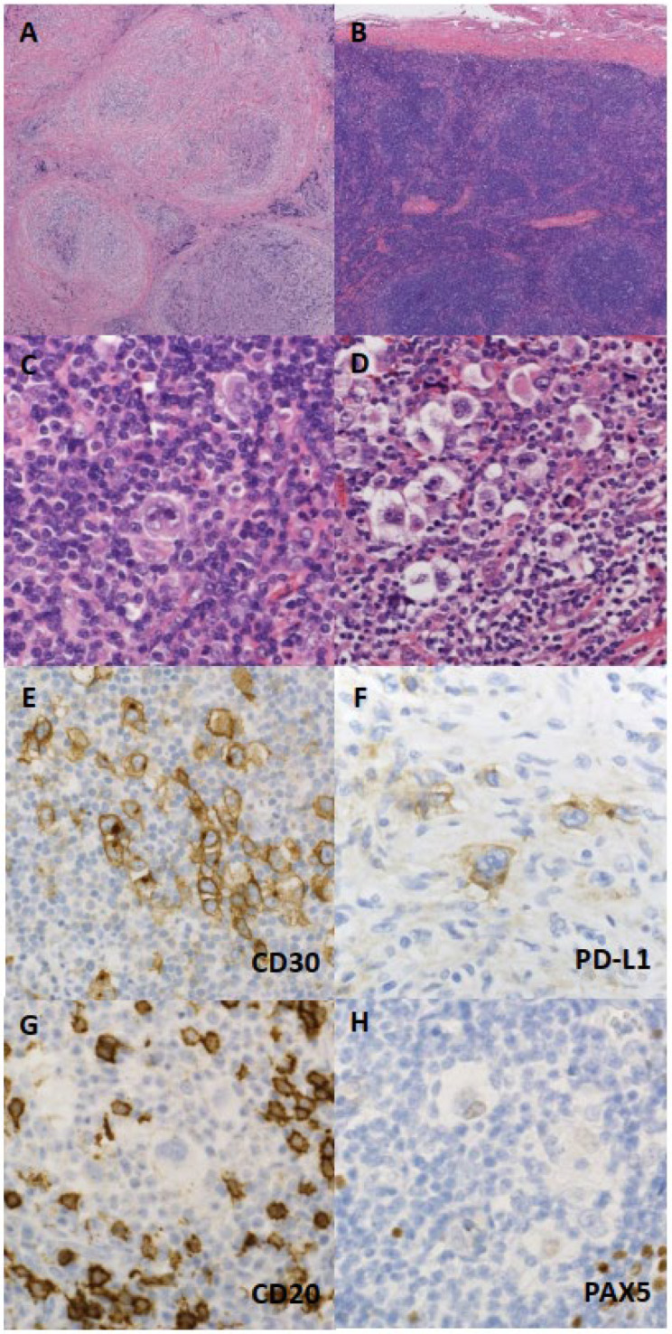
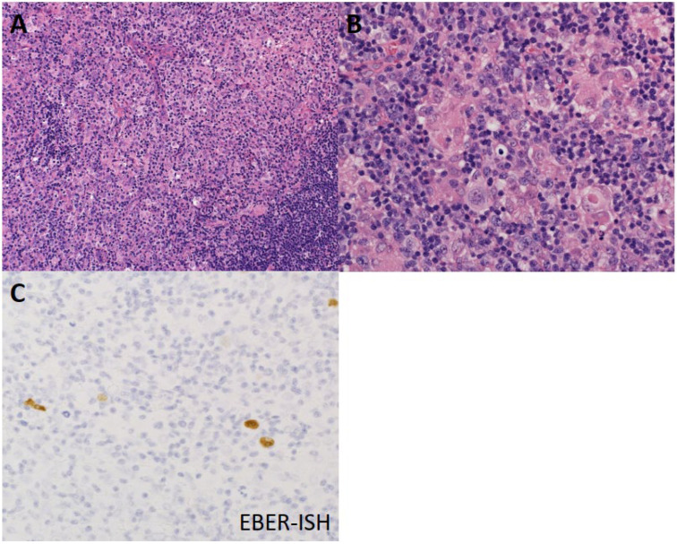
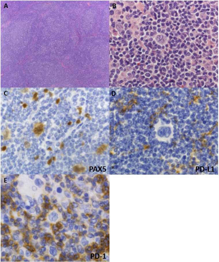
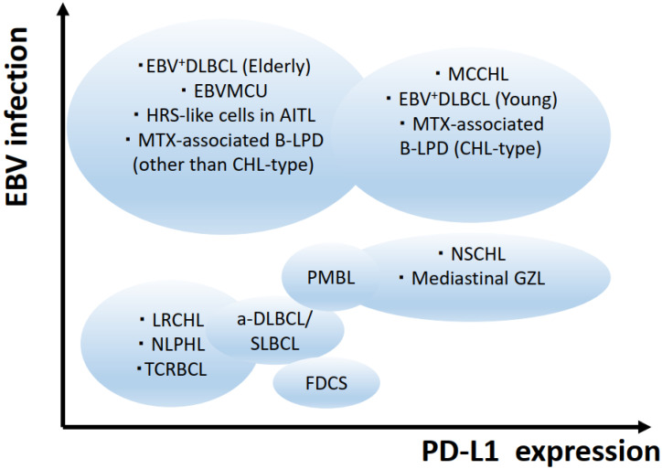
Hodgkin lymphomas (HLs) are lymphoid neoplasms that are morphologically defined as being composed of dysplastic cells, namely, Hodgkin and Reed-Sternberg cells, in a reactive inflammatory background. The biological nature of HLs has long been unclear; however, our understanding of HL-related genetics and tumor microenvironment interactions is rapidly expanding. For example, cell surface overexpression of programmed cell death 1 ligand 1 (CD274/PD-L1) is now considered a defining feature of an HL subset, and targeting such immune checkpoint molecules is a promising therapeutic option. Still, HLs comprise multiple disease subtypes, and some HL features may overlap with its morphological mimics, posing challenging diagnostic and therapeutic problems. In this review, we summarize the recent advances in understanding the biology of HLs, and discuss approaches to differentiating HL and its mimics.

Keywords: Hodgkin lymphoma; genetics; tumor microenvironment.


Conflict of interest statement

The authors declare no conflict of interest.

Figures

Figure 1
Figure 1
Histological and immunohistochemical features of nodular sclerosis classical Hodgkin lymphoma (NSCHL). (A) Cellular nodules are separated by collagen bundles. (B) Sclerotic thickening of the lymph node capsule is shown. (C) Neoplastic cells that are binucleated with a “mirror-image” appearance, called “Reed–Stenberg cells”. (D) Neoplastic cells with a lacuna-like space around the cytoplasm, called “lacunar cells”. (EH) Neoplastic cells expressing CD30 and PD-L1 (assessed using clone SP142), lacking CD20 expression, and with very weak PAX5 expression.
Figure 2
Figure 2
Histological features of mixed cellularity classical Hodgkin lymphoma (MCCHL). (A) Granuloma formation by histiocyte aggregation is observed in the background. (B) Neoplastic cells are scattered against a background rich in histiocytes and lymphocytes. (C) Neoplastic cells with highlighted Epstein–Barr virus RNA through in situ hybridization (EBER-ISH).
Figure 3
Figure 3
Histological and immunohistochemical features of nodular lymphocyte-predominant HL (NLPHL). (A) Vague nodular architecture is observed. (B) Scattered neoplastic cells feature polylobated (popcorn-like) nuclei, and are called lymphocyte-predominant (LP) cells. (C) LP cells show strong PAX5 expression. (D) LP cells lack PD-L1 expression (assessed using clone SP142). (E) LP cells are ringed by PD-1-positive “rosetting” T lymphocytes.
Figure 4
Figure 4
Images of the disease entities of classic Hodgkin lymphoma subtypes and its mimics. EBV = Epstein-Barr virus; AITL = angioimmunoblastic T-cell lymphoma; NSCHL = nodular sclerosis classic Hodgkin lymphoma; LRCHL = lymphocyte-rich classic Hodgkin lymphoma; MCCHL = mixed cellularity classic Hodgkin lymphoma; NLPHL = nodular lymphocyte-predominant Hodgkin lymphoma; DLBCL = diffuse large B-cell lymphoma; EBVMCU = EBV-positive mucocutaneous ulcer; MTX = methotrexate; LPD = lymphoproliferative disorder; PMBL = primary mediastinal large B-cell lymphoma; GZL = B-cell lymphoma unclassifiable, with features intermediate between DLBCL and CHL/Gray zone lymphoma; TCRBCL = T-cell/histiocyte-rich large B-cell lymphoma; a-DLBCL = anaplastic variant of DLBCL; SLBCL = sinusoidal large B-cell lymphoma; FDCS = follicular dendritic cell sarcoma.

Similar articles

Cited by

References

    1. Hodgkin T. On some Morbid Appearances of the Absorbent Glands and Spleen. Med. Chir. Trans. 1832;17:68–114. doi: 10.1177/095952873201700106. - DOI - PMC - PubMed
    1. Harris N.L., Jaffe E.S., Stein H., Banks P.M., Chan J.K., Cleary M.L., Delsol G., De Wolf-Peeters C., Falini B., Gatter K.C., et al. A revised European-American classification of lymphoid neoplasms: A proposal from the International Lymphoma Study Group. Blood. 1994;84:1361–1392. doi: 10.1182/blood.V84.5.1361.1361. - DOI - PubMed
    1. Campo E., Jaffe E.S., Cook J.R., Quintanilla-Martinez L., Swerdlow S.H., Anderson K.C., Brousset P., Cerroni L., de Leval L., Dirnhofer S., et al. The International Consensus Classification of Mature Lymphoid Neoplasms: A Report from the Clinical Advisory Committee. Blood. 2022 doi: 10.1182/blood.2022015851. - DOI - PMC - PubMed
    1. Swerdlow S.H. WHO Classification of Tumours of Haematopoietic and Lymphoid Tissues. 4th ed. Volume 2 International Agency for Research on Cancer; Lyon, France: 2017.
    1. Saito E., Matsuoka J. International comparison of Hodgkin and non-Hodgkin lymphoma incidence. Jpn. J. Clin. Oncol. 2020;50:96–97. doi: 10.1093/jjco/hyz202. - DOI - PubMed