. 2022 May 25;11(6):810.
doi: 10.3390/biology11060810.

Low-Salt Diet Reduces Anti-CTLA4 Mediated Systemic Immune-Related Adverse Events while Retaining Therapeutic Efficacy against Breast Cancer

Affiliations

Low-Salt Diet Reduces Anti-CTLA4 Mediated Systemic Immune-Related Adverse Events while Retaining Therapeutic Efficacy against Breast Cancer

Durga Khandekar et al. Biology (Basel). .

Abstract

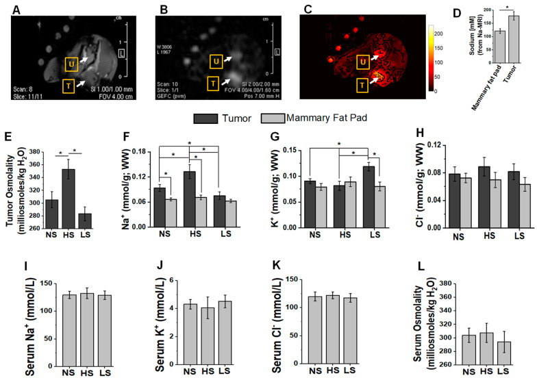
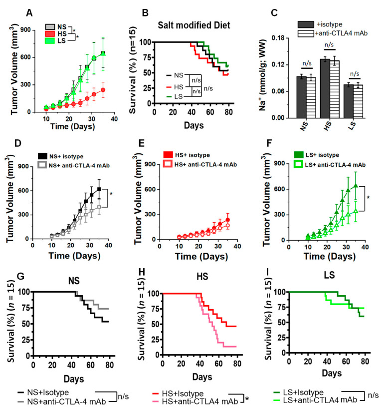
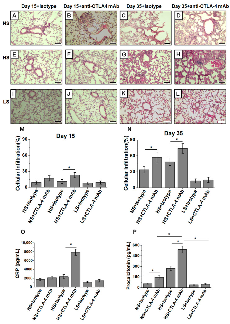
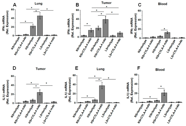
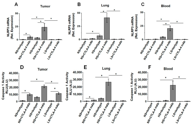
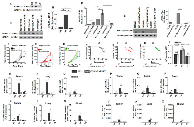
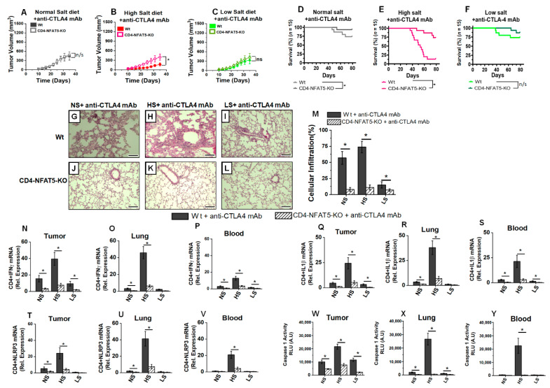
Immune checkpoint inhibitor (ICI) therapy has revolutionized the breast cancer treatment landscape. However, ICI-induced systemic inflammatory immune-related adverse events (irAE) remain a major clinical challenge. Previous studies in our laboratory and others have demonstrated that a high-salt (HS) diet induces inflammatory activation of CD4+T cells leading to anti-tumor responses. In our current communication, we analyzed the impact of dietary salt modification on therapeutic and systemic outcomes in breast-tumor-bearing mice following anti-cytotoxic T-lymphocyte-associated protein 4 (CTLA4) monoclonal antibody (mAb) based ICI therapy. As HS diet and anti-CTLA4 mAb both exert pro-inflammatory activation of CD4+T cells, we hypothesized that a combination of these would lead to enhanced irAE response, while low-salt (LS) diet through blunting peripheral inflammatory action of CD4+T cells would reduce irAE response. We utilized an orthotopic murine breast tumor model by injecting Py230 murine breast cancer cells into syngeneic C57Bl/6 mice. In an LS diet cohort, anti-CTLA4 mAb treatment significantly reduced tumor progression (day 35, 339 ± 121 mm3), as compared to isotype mAb (639 ± 163 mm3, p < 0.05). In an HS diet cohort, treatment with anti-CTLA4 reduced the survival rate (day 80, 2/15) compared to respective normal/regular salt (NS) diet cohort (8/15, p < 0.05). Further, HS plus anti-CTLA4 mAb caused an increased expression of inflammatory cytokines (IFNγ and IL-1β) in lung infiltrating and peripheral circulating CD4+T cells. This inflammatory activation of CD4+T cells in the HS plus anti-CTLA4 cohort was associated with the upregulation of inflammasome complex activity. However, an LS diet did not induce any significant irAE response in breast-tumor-bearing mice upon treatment with anti-CTLA4 mAb, thus suggesting the role of high-salt diet in irAE response. Importantly, CD4-specific knock out of osmosensitive transcription factor NFAT5 using CD4cre/creNFAT5flox/flox transgenic mice caused a downregulation of high-salt-mediated inflammatory activation of CD4+T cells and irAE response. Taken together, our data suggest that LS diet inhibits the anti-CTLA4 mAb-induced irAE response while retaining its anti-tumor efficacy.

Keywords: breast cancer; cancer biology; cytokines; immunotherapy; t-helper cells.


Conflict of interest statement

The authors declare no conflict of interest.

Figures

Figure 1
Figure 1
Sodium MRI in vivo live imaging of Py230-C57Bl/6 breast tumor. Py230 breast tumor cells were injected into right mammary fat pad in 8-week-old female C57Bl/6 mice. The imaging was performed on day 35 post-injection. (A) Proton-MRI; (B) Sodium(Na)-MRI; (C) heat map semi-quantitation of [Na+]. ‘T’ refers to orthotopic breast tumor; ‘U’ refers to urinary bladder (also serves as internal positive control as [Na+] is generally high in urine); (D) quantitative determination of [Na+] concentration from Na-MRI (n = 4). mean ± SEM, (*) p-value < 0.05, data analyzed by one-way ANOVA. Next, we determined tumor sodium accumulation following dietary salt (NaCl) modulation. (E) Tumor osmolality changes analyzed by vapor pressure osmometer following dietary salt modulation. (FH) Tumor and contralateral mammary fat pad [Na+], [K+], and [Cl] concentrations (mmol/g, WW; where WW refers to wet weight) following regular/normal-salt (NS, 1% NaCl), high-salt (HS, 4% NaCl) and low-salt (LS, 0.1% NaCl) diet; [Na+] and [K+] measured by flame atomic absorption, and [Cl] analyzed by silver nitrate titration. (IL) Plasma electrolyte and osmolality changes following dietary salt modification. (EL) Data analyzed by Tukey HSD pair-wise comparison and one-way ANOVA for multiple comparisons; data presented as mean ± SEM, n = 8 per cohort, (*) p-value < 0.05.
Figure 2
Figure 2
Tumor volume kinetics and survival analysis following salt-modified diet. (A) Tumor volume changes following dietary salt modification (NS, normal/regular salt; HS, high salt; LS, low salt); tumor volume kinetics followed for 5 weeks. Data presented as mean ± SEM, n = 8, statistical analysis was performed using two-way ANOVA multiple comparisons, Dunnet’s post-test (*) p-value < 0.05. (B) Survival rate in the three salt modified diet cohorts, tumor-bearing mice followed for 80 days. The n = 15 per cohort, statistical analysis performed by log-rank Mantel–Cox comparison. (C) [Na+] concentration in the murine breast tumors with isotype vs. anti-CTLA4 mAb treatment. Data analyzed by post hoc Bonferroni correction, n/s = not significant. (DF) Tumor progression kinetics in NS (D), HS (E) and LS (F) cohorts following treatment with anti-CTLA4 monoclonal antibody (mAb) or isotype control with 200 µg injected intraperitoneally (i.p.) on days 7, 10 and 13, and followed for 5 weeks. Data presented as mean ± SEM, n = 8, statistical analysis was performed using multiple t-test, (*) p-value < 0.05. (GI) Survival rate analysis in NS (G), HS (H) and LS (I) cohorts following treatment with anti-CTLA4 monoclonal antibody (mAb) or isotype control with 200 µg on days 7, 10 and 13, and followed for 80 days. The n = 15 per cohort, statistical analysis performed by log-rank Mantel–Cox comparison. Hazard ratio (HR) was performed by Mantel–Haenszel method.
Figure 3
Figure 3
Increased pneumonitis and lung infiltration in tumor-bearing mice on HS diet and treated with anti-CTLA4 mAb based ICI therapy. Lungs are harvested on days 15 and 35 from mice injected with Py230 tumor cells in the mammary fat pad flank region of C57Bl/6 mice and treated with either isotype or anti-CTLA4 mAb i.p. on days 7, 10 and 13. (AD) Tumor-bearing mice were fed on NS diet, lungs were harvested on day 15 (isotype, (A), and anti-CTLA4 mAb, (B)) and day 35 (isotype, (C), and anti-CTLA4 mAb, (D)). (EH) Tumor-bearing mice were fed on HS diet, lungs were harvested on day 15 (isotype, (E), and anti-CTLA4 mAb, (F)) and day 35 (isotype, (G), and anti-CTLA4 mAb, (H)). (IL) Tumor-bearing mice were fed on LS diet, lungs were harvested on day 15 (isotype, (I), and anti-CTLA4 mAb, (J)) and day 35 (isotype, (K), and anti-CTLA4 mAb, (L)). The slides were stained by H&E, and cell infiltration was measured under high power (40×) field. (M,N) Morphometric analysis for cellular infiltration on day 15 (M) and day 35 (N). The data are represented as a mean ± SEM for n = 6, with each slide read at 5 different high power fields and divided by 5. Scale bar = 200 μm. Statistical analysis performed by one-way ANOVA for multiple comparisons (*) p-value < 0.05. (O,P) Biochemical analysis of inflammatory markers, c-reactive protein (CRP) (O) and procalcitonin (P) in the blood from various experimental cohorts. Data analyzed by one-way ANOVA for multiple comparisons; data presented as mean ± SEM, n = 8 per cohort, (*) p-value < 0.05.
Figure 4
Figure 4
Low-salt diet reduces systemic irAE response in breast-tumor-bearing mice. The mRNA expression of inflammatory cytokines in CD4+T cells were analyzed in tumor-infiltrating, lung-infiltrating and peripheral circulation of breast tumor-bearing mice on ICI therapy following salt diet modification. (AC) The mRNA expression of IFNγ in CD4+T cells isolated from tumor (A), lung (B) and blood (C) in NS, HS and LS diet cohorts following treatment with anti-CTLA4 mAb or isotype control. (DF) The mRNA expression of IL-1β in CD4+T cells isolated from tumor (D), lung (E) and blood (F) in NS, HS and LS diet cohorts following treatment with anti-CTLA4 mAb or isotype control. Data analyzed by one-way ANOVA for multiple comparisons; data presented as mean ± SEM, n = 8 (biological replicates) per cohort, (*) p-value < 0.05.
Figure 5
Figure 5
Inflammasome activation in high-salt-diet-fed breast-tumor-bearing mice following anti-CTLA4 mAb therapy. (AC) The mRNA expression of NLRP3 in CD4+T cells isolated from tumor (A), lung (B) and blood (C) in NS, HS and LS diet cohorts following treatment with anti-CTLA4 mAb or isotype control. (DF) Proteolytic enzyme caspase-1 activity in CD4+T cells isolated from tumor (D), lung (E) and blood (F) in NS, HS and LS diet cohorts following treatment with anti-CTLA4 mAb or isotype control. Data analyzed by one-way ANOVA for multiple comparisons; data presented as mean ± SEM, n = 8 (biological replicates) per cohort, (*) p-value < 0.05.
Figure 6
Figure 6
NFAT5 is the upstream modulator of inflammasome activation in high-salt-mediated irAE response. (A) Western-blot-based protein analysis for NFAT5 expression in CD4+T cells isolated from tumor-infiltrating immune cells in NS, HS and LS diet cohorts. (B) qRT-PCR-based NFAT5 mRNA expression in CD4+T cells isolated from tumor-infiltrating immune cells in NS, HS and LS diet cohorts. (C,D) Western-blot- (C) and qRT-PCR- (D) based analysis of NFAT5 expression in CD4+T cells isolated from tumor-infiltrating immune cells in NS, HS and LS diet cohorts following treatment with anti-CTLA4 mAb or isotype control. (E) Transgenic murine model with CD4 specific NFAT5 knock out (CD4-NFAT5-KO) in C57Bl/6 background were utilized for further breast tumor studies. Western blot analysis confirmed the lack of NFAT5 expression in our CD4-NFAT5-KO model. (FH) Tumor progression kinetics in NS (F), HS (G) and LS (H) cohorts in wild-type (Wt) and CD4-NFAT5-KO transgenic mice injected with syngeneic Py230. Data presented as mean ± SEM, n = 8 (biological replicates); statistical analysis was performed using multiple t-test, (*) p-value < 0.05; n/s = not significant. (IK) Survival rate in the three salt-modified diet cohorts—NS (I), HS (J) and LS (K)—in breast-tumor-bearing Wt and CD4-NFAT5-KO mice followed for 80 days. The n =15 per cohort, statistical analysis performed by log-rank Mantel–Cox comparison. (L) CD4-specific knock out of NFAT5 did not change the tumor [Na+] accumulation. (MO) The mRNA expression of IFNγ in CD4+T cells isolated from tumor (M), lung (N) and blood (O) of Wt and CD4-NFAT5-KO in NS, HS and LS diet cohorts. (PR) The mRNA expression of IL1β in CD4+T cells isolated from tumor (P), lung (Q) and blood (R) of Wt and CD4-NFAT5-KO in NS, HS and LS diet cohorts. (SU) The mRNA expression of NLRP3 in CD4+T cells isolated from tumor (S), lung (T) and blood (U) of Wt and CD4-NFAT5-KO in NS, HS and LS diet cohorts. (VX) Caspase-1 activity CD4+T cells in tumor (V), lung (W) and blood (X) from Wt and CD4-NFAT5-KO in NS, HS and LS diet cohorts. Data analyzed by one-way ANOVA for multiple comparisons; data presented as mean ± SEM, n = 8 (biological replicates) per cohort, (*) p-value < 0.05.
Figure 7
Figure 7
NFAT5 knock out reduces high-salt-mediated inflammatory activation of CD4+T cells and systemic inflammatory response in breast-tumor-bearing mice. (AC) Tumor progression kinetics in NS (A), HS (B) and LS (C) cohorts in wild-type (Wt) and CD4-NFAT5-KO transgenic mice injected with syngeneic Py230 following treatment with anti-CTLA4 mAb. Data presented as mean ± SEM, n = 8, statistical analysis was performed using multiple t-test, (*) p-value < 0.05, n/s = not significant. (DF) Survival rate in the three salt-modified diet cohorts—NS (D), HS (E) and LS (F), in breast tumor-bearing Wt and CD4-NFAT5-KO mice following treatment with anti-CTLA4 mAb. The n =15 per cohort, statistical analysis performed by log-rank Mantel–Cox comparison. (GL) Tumor-bearing mice were fed on NS (G,J), HS (H,K) and LS (I,L) diet in breast-tumor-bearing Wt and CD4-NFAT5-KO mice; lungs were harvested on day 35 and analyzed by H&E staining for cellular infiltration. (M) Quantitative morphometric analysis of the cellular infiltration in lungs harvested on day 35 from Wt and CD4-NFAT5-KO tumor-bearing mice following treatment with anti-CTLA4 mAb. The data are represented as mean ± SEM for n = 6; scale bar = 200 μm. (GM), with each slide read at 5 different high-power fields and divided by 5. Statistical analysis performed by one-way ANOVA for multiple comparisons (*) p-value < 0.05. (NP) The mRNA expression of IFNγ in CD4+T cells isolated from tumor (N), lung (O) and blood (P) of Wt and CD4-NFAT5-KO in NS, HS and LS diet cohorts following treatment with anti-CTLA4 mAb. (QS) The mRNA expression of IL1β in CD4+T cells isolated from tumor (Q), lung (R) and blood (S) of Wt and CD4-NFAT5-KO in NS, HS and LS diet cohorts following treatment with anti-CTLA4 mAb. (TV) The mRNA expression of NLRP3 in CD4+T cells isolated from tumor (T), lung (U) and blood (V) of Wt and CD4-NFAT5-KO in NS, HS and LS diet cohorts following treatment with anti-CTLA4 mAb. (WY) Caspase-1 activity CD4+T cells in tumor (W), lung (X) and blood (Y) from Wt and CD4-NFAT5-KO in NS, HS and LS diet cohorts following treatment with anti-CTLA4 mAb. Data analyzed by one-way ANOVA for multiple comparisons; data presented as mean ± SEM, n = 8 (biological replicates) per cohort (NY), (*) p-value < 0.05.

Similar articles

Cited by

References

    1. Robert C. A decade of immune-checkpoint inhibitors in cancer therapy. Nat Commun. 2020;11:3801. doi: 10.1038/s41467-020-17670-y. - DOI - PMC - PubMed
    1. American Cancer Society . Cancer Facts & Figures (2022) American Cancer Society; Atlanta, GA, USA: 2022.
    1. Thomas R., Al-Khadairi G., Decock J. Immune Checkpoint Inhibitors in Triple Negative Breast Cancer Treatment: Promising Future Prospects. Front. Oncol. 2021;10:600573. doi: 10.3389/fonc.2020.600573. - DOI - PMC - PubMed
    1. Zheng M. Tumor mutation burden for predicting immune checkpoint blockade response: The more, the better. J. Immunother. Cancer. 2022;10:e003087. doi: 10.1136/jitc-2021-003087. - DOI - PMC - PubMed
    1. Sivapiragasam A., Ashok Kumar P., Sokol E.S., Albacker L.A., Killian J.K., Ramkissoon S.H., Huang R.S.P., Severson E.A., Brown C.A., Danziger N., et al. Predictive Biomarkers for Im mune Checkpoint Inhibitors in Metastatic Breast Cancer. Cancer Med. 2021;10:53–61. doi: 10.1002/cam4.3550. - DOI - PMC - PubMed