. 2022 May 28;11(6):831.
doi: 10.3390/biology11060831.

Community Vertical Composition of the Laguna Negra Hypersaline Microbial Mat, Puna Region (Argentinean Andes)

Affiliations

Community Vertical Composition of the Laguna Negra Hypersaline Microbial Mat, Puna Region (Argentinean Andes)

Flavia Jaquelina Boidi et al. Biology (Basel). .

Abstract

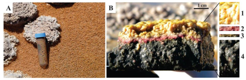
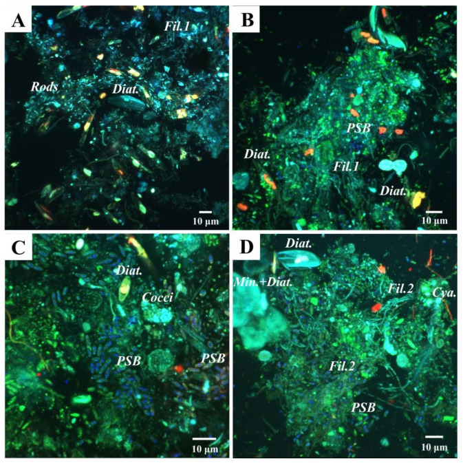
The Altiplano-Puna region is a high-altitude plateau in South America characterized by extreme conditions, including the highest UV incidence on Earth. The Laguna Negra is a hypersaline lake located in the Catamarca Province, northwestern Argentina, where stromatolites and other microbialites are found, and where life is mostly restricted to microbial mats. In this study, a particular microbial mat that covers the shore of the lake was explored, to unravel its layer-by-layer vertical structure in response to the environmental stressors therein. Microbial community composition was assessed by high-throughput 16S rRNA gene sequencing and pigment content analyses, complemented with microscopy tools to characterize its spatial arrangement within the mat. The top layer of the mat has a remarkable UV-tolerance feature, characterized by the presence of Deinococcus-Thermus and deinoxanthin, which might reflect a shielding strategy to cope with high UV radiation. Chloroflexi and Deltaproteobacteria were abundant in the second and third underlying layers, respectively. The bottom layer harbors copious Halanaerobiaeota. Subspherical aggregates composed of calcite, extracellular polymeric substances, abundant diatoms, and other microorganisms were observed all along the mat as the main structural component. This detailed study provides insights into the strategies of microbial communities to thrive under high UV radiation and hypersalinity in high-altitude lakes in the Altiplano-Puna region.

Keywords: Andes; Puna region; UV radiation; extreme environment; high-altitude; hypersaline lake; microbial diversity; pigments.


Conflict of interest statement

The authors declare no conflict of interest.

Figures

Figure 1
Figure 1
Geographical location of the studied area. (A) Argentina map showing the Catamarca Province in yellow; (B) Catamarca Province map showing the location of the Laguna Verde Saline Complex (red square); (C) Laguna Negra satellite image showing the study area location (red square); (D) Overview of the “Stromatolite Belt” in the southeastern shore of the Laguna Negra, where microbialites and microbial mats occur; (E) A detailed image of microbial mats surrounding oncoids in the “Stromatolite Belt”.
Figure 2
Figure 2
Studied microbial mat. (A) Overview; (B) Vertical section of one portion of the microbial mat showing the four dissected layers: pink-orange Layer 1, purple Layer 2, green Layer 3, and black Layer 4.
Figure 3
Figure 3
Alpha diversity metrics of the four layers studied. Sampling effort was normalized at 3250 reads per sample. ASVs: amplicon sequence variants.
Figure 4
Figure 4
Relative abundance of bacterial phyla and Archaea in the Laguna Negra microbial mat layers. Assignations are derived from sequences analysis of the V4 hypervariable region of 16S rRNA gene. “Minor phyla” indicates less than 1% of abundance and included Acetothermia, Acidobacteria, Chlamydiae, Cloacimonetes, Firmicutes, Fusobacteria, Marinimicrobia (SAR406 clade), Omnitrophicaeota, and WS1. “Bacteria; Other” indicates unclassified.
Figure 5
Figure 5
Venn diagram showing the number of unique or shared ASVs between the layers of the microbial mat.
Figure 6
Figure 6
Relative abundance and taxonomic assignment of the main families found in each layer. The figure shows families with relative abundances higher than 1% of overall dataset.
Figure 7
Figure 7
Total absorbance spectra of the pigments extracted with methanol from the four layers individually.
Figure 8
Figure 8
SEM images of the studied microbial mat layers. (A) Abundant pennate diatoms (image from Layer 1); (B) Irregular-subspherical diatom-microbe-mineral aggregates (image from Layer 2); (C,D) Filamentous and rod-shaped microbes within the EPS matrix together with mineral particles; (E) Diatoms and microbes immersed within the EPS matrix where clusters of irregular-globular mineral particles are also observed (blue arrow). Note the fibrillar textures of EPS (red arrow); (F) Subspherical aggregates where diatoms, EPS, and mineral remains are observed (picture from Layer 3); (G) Irregularly shaped mineral cluster where subhedral carbonate mineral particles are observed (image from Layer 4).
Figure 9
Figure 9
Confocal Laser Scanning Microscopy (CLSM) images of the studied microbial mat layers. (A,B) Top layer of the mat, where (A) shows a fixed sample without staining and (B) shows a sample stained with Syto®9 (DNA staining). Diatoms are visible in light blue (Diat.), and also rod-shaped bacteria (Rods) and filamentous bacteria (Fil.1). Purple Sulfur Bacteria (PSB) are detected due to laser reflection of the sulfur globules inside the cells in dark blue; (C,D) CLSM images that correspond to Layers 2 and 3, both stained with Syto®9; (C) Abundant PSB are visualized, as well as dense colonies of autoflourescent cocci (Cocci), and some diatoms (Diat.); (D) Filamentous bacteria, possibly Chloroflexi (Fil.2), diatoms (Diat.), and diatoms frustules associated with globules of minerals (Min. + Diat.) detected in light blue, PSB in dark blue, and a few Cyanobacteria (Cya.) in red, due to chlorophyll a fluorescence.

Similar articles

Cited by

References

    1. Franks J., Stolz J.F. Flat laminated microbial mat communities. Earth-Sci. Rev. 2009;96:163–172. doi: 10.1016/j.earscirev.2008.10.004. - DOI
    1. Gerdes G. What Are Microbial Mats? In: Seckbach J., Oren A., editors. Microbial Mats: Modern and Ancient Microorganisms in Stratified Systems. 1st ed. Springer Science & Business Media B.V.; Dordrecht, The Netherlands: 2010. pp. 5–25.
    1. Prieto-Barajas C.M., Valencia-Cantero E., Santoyo G. Microbial mat ecosystems: Structure types, functional diversity, and biotechnological application. Electron. J. Biotechnol. 2018;31:48–56. doi: 10.1016/j.ejbt.2017.11.001. - DOI
    1. Marais D.J.D. Microbial mats and the early evolution of life. Trends Ecol. Evol. 1990;5:140–144. doi: 10.1016/0169-5347(90)90219-4. - DOI - PubMed
    1. Seckbach J., Oren A. Microbial Mats: Modern and Ancient Microorganisms in Stratified Systems. 1st ed. Springer Science & Business Media B.V.; Dordrecht, The Netherlands: 2010. pp. 389–539.

LinkOut - more resources