. 2022 Jun 15;12(6):789.
doi: 10.3390/brainsci12060789.

Synaptic Dysfunction by Mutations in GRIN2B: Influence of Triheteromeric NMDA Receptors on Gain-of-Function and Loss-of-Function Mutant Classification

Affiliations

Synaptic Dysfunction by Mutations in GRIN2B: Influence of Triheteromeric NMDA Receptors on Gain-of-Function and Loss-of-Function Mutant Classification

Marwa Elmasri et al. Brain Sci. .

Abstract

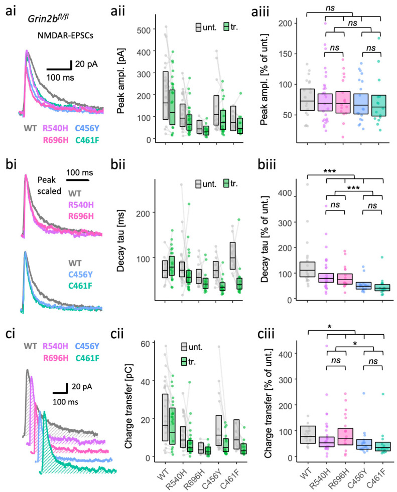
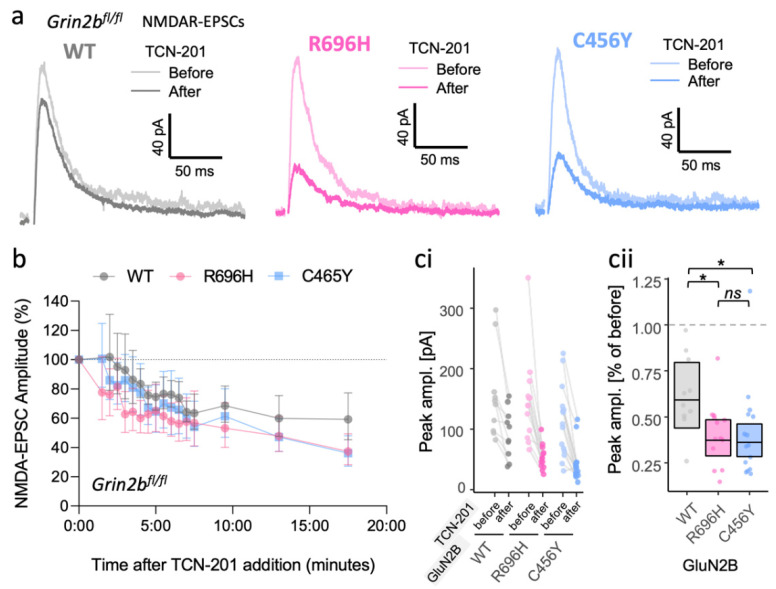
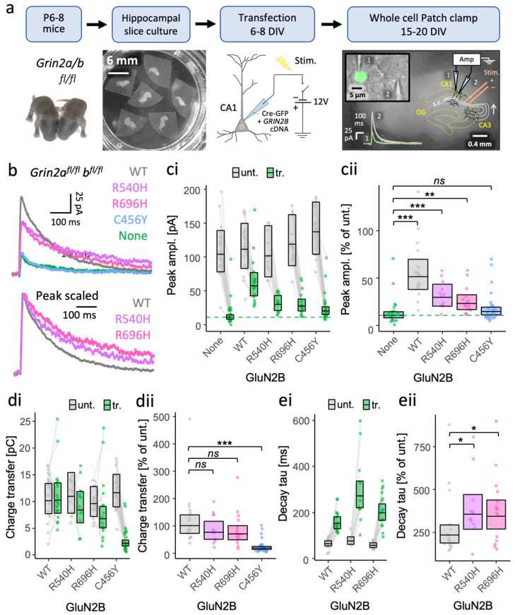
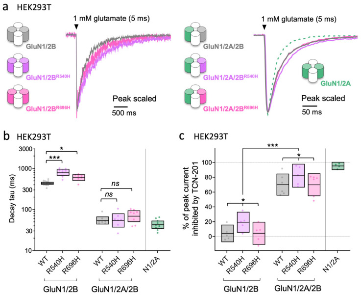
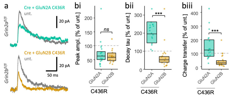
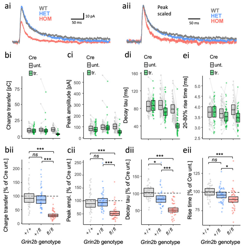
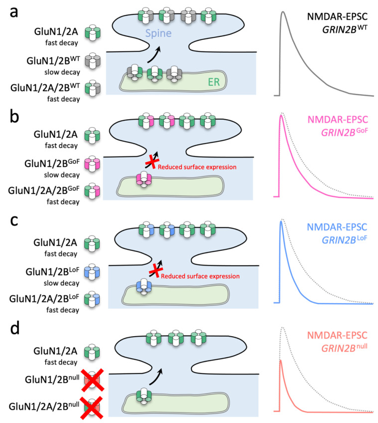
GRIN2B mutations are rare but often associated with patients having severe neurodevelopmental disorders with varying range of symptoms such as intellectual disability, developmental delay and epilepsy. Patient symptoms likely arise from mutations disturbing the role that the encoded NMDA receptor subunit, GluN2B, plays at neuronal connections in the developing nervous system. In this study, we investigated the cell-autonomous effects of putative gain- (GoF) and loss-of-function (LoF) missense GRIN2B mutations on excitatory synapses onto CA1 pyramidal neurons in organotypic hippocampal slices. In the absence of both native GluN2A and GluN2B subunits, functional incorporation into synaptic NMDA receptors was attenuated for GoF mutants, or almost eliminated for LoF GluN2B mutants. NMDA-receptor-mediated excitatory postsynaptic currents (NMDA-EPSCs) from synaptic GoF GluN1/2B receptors had prolonged decays consistent with their functional classification. Nonetheless, in the presence of native GluN2A, molecular replacement of native GluN2B with GoF and LoF GluN2B mutants all led to similar functional incorporation into synaptic receptors, more rapidly decaying NMDA-EPSCs and greater inhibition by TCN-201, a selective antagonist for GluN2A-containing NMDA receptors. Mechanistic insight was gained from experiments in HEK293T cells, which revealed that GluN2B GoF mutants slowed deactivation in diheteromeric GluN1/2B, but not triheteromeric GluN1/2A/2B receptors. We also show that a disease-associated missense mutation, which severely affects surface expression, causes opposing effects on NMDA-EPSC decay and charge transfer when introduced into GluN2A or GluN2B. Finally, we show that having a single null Grin2b allele has only a modest effect on NMDA-EPSC decay kinetics. Our results demonstrate that functional incorporation of GoF and LoF GluN2B mutants into synaptic receptors and the effects on EPSC decay times are highly dependent on the presence of triheteromeric GluN1/2A/2B NMDA receptors, thereby influencing the functional classification of NMDA receptor variants as GoF or LoF mutations. These findings highlight the complexity of interpreting effects of disease-causing NMDA receptor missense mutations in the context of neuronal function.

Keywords: central nervous system; de novo mutations; electrophysiology; ionotropic glutamate receptors; synaptic transmission.


Conflict of interest statement

The authors declare no conflict of interest. The funders had no role in the design of the study; in the collection, analyses, or interpretation of data; in the writing of the manuscript, or in the decision to publish the results.

Figures

Figure 2
Figure 2
Gain- and loss-of-function GluN2B mutants are both associated with more rapidly decaying NMDA-EPSCs in GluN2B knockout neurons. NMDA-EPSC+20 mV peak amplitudes (a), decay time constants (b) or charge transfer (c) in Grin2bfl/fl (untransfected) neurons and Grin2b−/− neurons rescued with human GluN2B WT, GoF (R540H and R696H) or LoF (C456Y or C461F) mutants (transfected). (ac) (i) Representative NMDA-EPSCs (average of 10 sweeps) from transfected CA1 neurons; (ii) data points of measurements made in individual neurons, matched data points, for simultaneously recorded untransfected and transfected neurons, are connected by a line; (iii) response ratios (transfected/untransfected) are expressed as a percentage and plotted for each pair of transfected–untransfected neurons. Crossbars in (ii) and (iii) show the estimated marginal means with 95% confidence intervals backtransformed from the linear mixed models (Figure S1.4 and S1.6–7). Hypothesis tests are orthogonal contrasts based on a priori classification of the mutations (see Table S1). Standardised effect sizes (r) for comparisons of each mutant with WT for response ratios of: (aiii) peak amplitudes were −0.02, −0.04, −0.06, and −0.08; (biii) decay taus were −0.19, −0.22, −0.42, and −0.47; and (ciii) charge transfer was −0.09, −0.04, −0.24, and −0.31, for mutants R540H, R696H, C456Y, and C461F, respectively (N = 95). ns = not significant (at α = 0.05), * = p < 0.05, *** = p < 0.001.
Figure 3
Figure 3
Gain- and loss-of-function GluN2B mutants both increase inhibition of NMDA-EPSCs in GluN2B knockout neurons by the GluN2A-selective antagonist TCN-201. (a) Representative pharmacologically isolated NMDA-EPSCs (average of 20 sweeps) from transfected CA1 neurons before and 17.5 min after application of 10 μM TCN-201. (b) NMDA-EPSC+20 mV peak amplitudes expressed as a % of the initial EPSC size (before application of 10 μM TCN-201) and summarised for all time points as the geometric mean and 95% confidence intervals. (ci) NMDA-EPSC peak amplitudes before and 17.5 min after the addition of 10 µM TCN-201; each point represents the peak of the ensemble mean of 20 NMDA-EPSCs, each pair of points connected by a line corresponds to measurements made from individual neurons before and after addition of TCN-201; (cii) response ratios (transfected/untransfected) calculated from (ci) are expressed as a percentage and plotted for each pair of before–after recordings. Crossbars show the estimated marginal means with 95% confidence intervals backtransformed from the linear mixed models (Figure S1.8). Standardised effect sizes (r) for each mutant compared to WT were −0.37 and −0.40 for mutants R696H and C456Y, respectively (N = 38). ns = not significant (at α = 0.05), * = p < 0.05.
Figure 1
Figure 1
More effective rescue of NMDA-EPSCs in GluN2A/B double knockout neurons by putative GoF mutants (R696H and R540H) than by LoF mutation (C456Y). (a) Experimental protocol used to test functional incorporation of GluN2B mutants. Plasmids expressing Cre-GFP and human GRIN2B cDNA were cotransfected into CA1 pyramidal neurons of organotypic hippocampal slices from Grin2afl/flbfl/fl mice. With transfection, the action of Cre-GFP at floxed Grin2a and Grin2b alleles knocks out native mouse GluN2A and 2B protein expression concurrently with expressing human GluN2B variants (WT or mutant). Untransfected neurons continue to express native NMDA receptors (e.g., GluN2A and GluN2B). (b) Representative NMDA-EPSC+20 mV (average of 30 sweeps) from transfected CA1 neurons. (c,d) NMDA-EPSC+20 mV peak amplitudes and charge transfer, respectively, for Grin2afl/flbfl/fl (untransfected, abbrv. Unt.) neurons, and Grin2a−/−b−/− neurons rescued with human GluN2B WT, GoF (R540H, R696H) or LoF (C456Y) mutants (transfected, abbrv. Tr.). (e) NMDA-EPSC+20 mV decay time constant (tau) and charge transfer, respectively, for Grin2afl/flbfl/fl neurons, and Grin2a−/−b−/− neurons rescued with human GluN2B WT, and GoF mutants R540H and R696H; (ce) (i) data points of measurements made in individual neurons; matched data points, for simultaneously recorded untransfected (unt.) and transfected (tr.) neurons, are connected by a line; (ii) response ratios (transfected/untransfected) are expressed as a percentage and plotted for each pair of transfected-untransfected neurons. Crossbars in (i) and (ii) show the estimated marginal means with 95% confidence intervals backtransformed from the linear mixed models (Figure S1.1–1.3 for (ce) respectively). Post hoc pairwise comparisons were made using Dunnett’s stepdown procedure. Standardised effect sizes (r) for comparisons of response ratios for: (cii) peak amplitudes (vs control: no GRIN2B cDNA, a.k.a. None) were +0.62, +0.39, +0.32 and +0.10 for WT, R540H, R696H, and C456Y, respectively (N = 90); or (dii) charge transfer (vs control: WT) was −0.12, −0.17 and −0.68 for R540H, R696H, and C456Y, respectively (N = 71) of (eii); decay time constant (tau) was +0.33 and +0.33 for mutants R540H and R696H, respectively (N = 47). Data for the NMDA-EPSC amplitudes in Grin2a−/−b−/− neurons with and without WT GluN2B were published in [40]. ns = not significant (at α = 0.05), * = p < 0.05, ** = p < 0.01, *** = p < 0.001.
Figure 4
Figure 4
Assembly of mutant GluN2B with GluN2A in triheteromeric GluN1/2A/2B receptors can abate functional effects of GoF missense GluN2B variants. (a) Representative fast-application whole-cell patch-clamp recordings of NMDA receptor responses normalized to the peak amplitude. The NMDA receptor subtypes are expressed in HEK293T cells and responses are activated by brief 5 ms exposures to 1 mM glutamate in the continuous presence of 100 μM glycine. (b) Decay time constants for WT and mutant NMDA receptor subtypes. Crossbars represent the geometric mean and 95% confidence intervals. The statistical comparisons presented are Dunnett’s post hoc test results following a significant mutation x subtype interaction (F (2,43) = 6.66, p = 0.003) from a two-way ANOVA (Type III) on log10-transformed decay tau values. (c) Inhibition by 5 µM TCN-201 of response amplitudes from WT and mutant NMDA receptor subtypes activated brief 5 ms application of 1 mM glutamate in the continuous presence of 3 μM glycine. Crossbars represent the mean and 95% confidence intervals. The statistical results presented in (c) are for the main effects of mutation (F (2,26) = 3.91, p = 0.033) and subtype (F (1,26) = 206.6, p ≤ 0.001) from a two-way ANOVA (Type III) on the % of peak current inhibited by TCN-201; the mutation x subtype interaction was not statistically significant (F (2,26) = 0.045, p = 0.96). In both (b) and (c), factor levels for mutation were WT, R540H and R696H; factor levels for subtype were diheteromer (GluN1/2B) and triheteromer (GluN1/2A/2B). ns = not significant (at α = 0.05), * = p < 0.05, *** = p < 0.001.
Figure 5
Figure 5
Subunit-dependent outcome of identical disease-associated LoF mutations in GRIN2A and GRIN2B. (a) Representative NMDA-EPSCs (average of 10 sweeps) from Grin2bfl/fl (untransfected abbrv. unt.) neurons, and Grin2b−/− neurons rescued with human GluN2AC436R or GluN2BC436R. (b) Ratios (transfected/untransfected) of the response (peak amplitude, decay time constant or charge transfer) are expressed as a percentage and plotted for each pair of transfected-untransfected neurons. Crossbars in (ii) and (iii) show the estimated marginal means with 95% confidence intervals backtransformed from the linear mixed models (LMM). The p-values from the hypothesis tests are derived from the same LMM (Figures S1.9–S1.11). Standardised effect sizes (r) for GluN2 subunit background (of the C436R mutation) on response ratios were (bi) for peak amplitudes, −0.08; (bii) for decay taus, −0.75; and (biii) for charge transfer, −0.62 (N = 33). ns = not significant (at α = 0.05), *** = p < 0.001.
Figure 6
Figure 6
Dose-dependent effects of Grin2b null alleles on NMDA-EPSC kinetics. (a) Representative NMDA-EPSCs from Grin2b+/+, Grin2b+/− or Grin2b−/− neurons. NMDA-EPSC+20 mV charge transfer (b), peak amplitude (c), decay time constant (d) and 20–80% rise-time (e) in Grin2b+/+, Grin2b+/fl or Grin2bfl/fl (untransfected) neurons and Grin2b+/+, Grin2b+/− or Grin2b−/− neurons (transfected, with Cre-GFP). (be) (i) Data points of measurements made in individual neurons. Matched data points, for simultaneously recorded untransfected and transfected neurons are connected by a line; (ii) response ratios (transfected/untransfected) are expressed as a percentage and plotted for each pair of transfected-untransfected neurons. Crossbars in (i) and (ii) show the estimated marginal means with 95% confidence intervals backtransformed from the fitted linear mixed models (Figure S1.12–S1.15). Post hoc pairwise comparisons were made according to the Westfall stepwise procedure. Standardised effect sizes (r) for comparisons of each genotype with WT for response ratios, for heterozygous and homozygous genotypes, respectively, were (aii) for charge transfer, −0.05 and −0.69; (bii) for peak amplitude, +0.04 and −0.43; (cii) for decay taus, −0.24 and −0.63; and (dii) for rise-time, −0.14 and −0.33, (N = 107). ns = not significant (at α = 0.05), * = p < 0.05, *** = p < 0.001.
Figure 7
Figure 7
Scheme summarizing how different molecular defects in the agonist binding domain of GluN2B converge to accelerate NMDA-EPSCs in CA1 neurons. (a) Schaffer collateral synapses onto CA1 neurons contain triheteromeric receptors (GluN1/2A/2B) and a small population of diheteromeric receptors (GluN1/2A and GluN1/2B). (b) GoF missense mutations in GluN2B cause GluN1/2B receptors to have more prolonged decay (Figure 1b,e), but do not traffic effectively to synapses (Figure 1b,c). Synaptic GluN1/2A/2B receptors with GoF GluN2B mutations traffic comparatively better than their GluN1/2B counterparts (Figure 2a), but the NMDA-EPSC time course is accelerated owing to the dominance of GluN2A on the deactivation of triheteromeric NMDA receptors (Figure 4b). (c) LoF missense mutations (that strongly reduce GluN2B surface expression) lead to an absence of mutant GluN1/2B receptors (Figure 1b,c), and likely also a greater representation of GluN1/2A receptors at synapses (Figure 2a). (d) Genetic deletion (i.e., the null allele) of Grin2b prevents the formation of NMDA receptors with any GluN2B subunits. GluN2A cannot fully compensate for the loss of (both) Grin2b alleles (Figure 6b). Since synapses in neurons with either GoF or LoF GluN2B missense mutations have fewer GluN1/2B diheteromers, they both exhibit more extensive inhibition of their associated NMDA-EPSCs by TCN-201 (compared to WT) (Figure 3).

Similar articles

Cited by

References

    1. Paoletti P., Bellone C., Zhou Q. NMDA Receptor Subunit Diversity: Impact on Receptor Properties, Synaptic Plasticity and Disease. Nat. Rev. Neurosci. 2013;14:383–400. doi: 10.1038/nrn3504. - DOI - PubMed
    1. Hansen K.B., Wollmuth L.P., Bowie D., Furukawa H., Menniti F.S., Sobolevsky A.I., Swanson G.T., Swanger S.A., Greger I.H., Nakagawa T., et al. Structure, Function, and Pharmacology of Glutamate Receptor Ion Channels. Pharmacol. Rev. 2021;73:298–487. doi: 10.1124/pharmrev.120.000131. - DOI - PMC - PubMed
    1. Kew J.N., Koester A., Moreau J.L., Jenck F., Ouagazzal A.M., Mutel V., Richards J.G., Trube G., Fischer G., Montkowski A., et al. Functional Consequences of Reduction in NMDA Receptor Glycine Affinity in Mice Carrying Targeted Point Mutations in the Glycine Binding Site. J. Neurosci. 2000;20:4037–4049. doi: 10.1523/JNEUROSCI.20-11-04037.2000. - DOI - PMC - PubMed
    1. Kleckner N.W., Dingledine R. Requirement for Glycine in Activation of NMDA-Receptors Expressed in Xenopus Oocytes. Science. 1988;241:835–837. doi: 10.1126/science.2841759. - DOI - PubMed
    1. Mothet J.-P., Parent A.T., Wolosker H., Brady R.O., Linden D.J., Ferris C.D., Rogawski M.A., Snyder S.H. D-Serine Is an Endogenous Ligand for the Glycine Site of the N-Methyl-d-Aspartate Receptor. Proc. Natl. Acad. Sci. USA. 2000;97:4926–4931. doi: 10.1073/pnas.97.9.4926. - DOI - PMC - PubMed

LinkOut - more resources