Case Reports
. 2022 Jun 19;12(6):802.
doi: 10.3390/brainsci12060802.

High Myoinositol on Proton MR Spectroscopy Could Be a Potential Signature of Papillary Tumors of the Pineal Region-Case Report of Two Patients

Affiliations
Case Reports

High Myoinositol on Proton MR Spectroscopy Could Be a Potential Signature of Papillary Tumors of the Pineal Region-Case Report of Two Patients

Albert Pons-Escoda et al. Brain Sci. .

Abstract

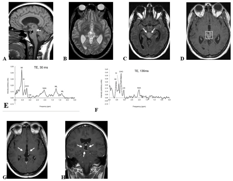
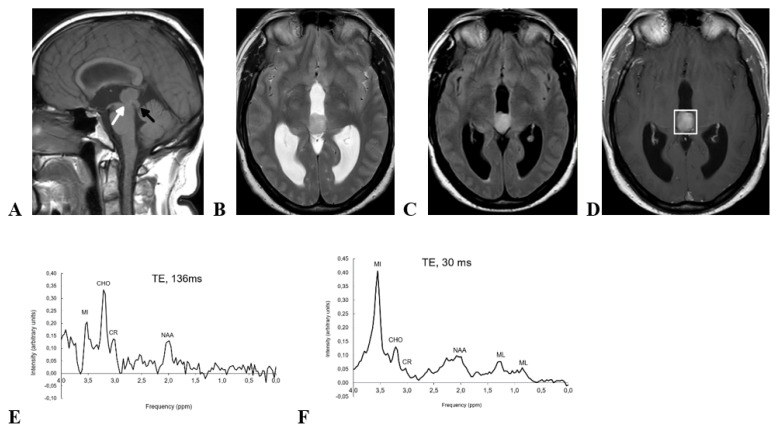
Papillary tumor of the pineal region (PTPR) is an uncommon entity in which a presurgical suspicion may be crucial for patient management. Maximal safe neurosurgical resection is of choice when PTPR is suspected, whereas non-surgical approaches can be considered in other tumors of the pineal region, such as pineocytoma or concrete subtypes of germ-cell tumors. In general terms, imaging features of tumors of the pineal region have been reported to be unspecific. Nevertheless, in this report, we describe two pathology-confirmed PTPRs in which presurgical proton MR spectroscopy demonstrated extremely high myoinositol, a pattern which drastically differs from that of other pineal tumors. We hypothesize that this high myoinositol may be related to PTPR's known ependymal component, and that it could be used as a specific non-invasive diagnostic signature.

Keywords: NMR spectroscopy; differential diagnosis; myoinositol; pineal neoplasms.


Conflict of interest statement

The authors declare no conflict of interest.

Figures

Figure 1
Figure 1
Preoperative MRI (AD), preoperative 1H-MRS (E,F), and postoperative MRI (G,H) of a 30-yr-old woman with pathology-confirmed PTPR. (A) Sagital T1-wi show an inhomogeneous pineal region mass with small areas of cystic degeneration. Sylvian aqueduct compression secondary to the mass (black arrow) and midbrain tegmentum infiltration (white arrow, see also Figure 1C) are shown in the image. (B) Axial T2-wi show that the mass signal is heterogeneous with multiple small cystic areas. Tumor margins are well defined at this level, and slight mass-effect is produced to both thalami. (C) Axial FLAIR-wi at a lower level than B shows midbrain infiltration (white arrows). (D) Contrast-enhanced T1-wi confirms a well-defined mass at this level with small cystic areas and non-homogeneous enhancement. Voxel for spectroscopy is shown. (E) Short TE 1H-MRS (TE, 30 ms) shows very high myoinositol signal at 3.55 ppm. (F) Long TE 1H-MRS (TE, 136 ms) shows that the intensity at 3.55 ppm is reduced at this echo time due to short T2 relaxation time of myoinositol. MI, myoinositol; SI, scyllo-inositol; CHO, choline; CR, creatine; NAA, N-acetyl-aspartate; ML, mobile lipids. (G,H) Postoperative axial and coronal contrast-enhanced T1-wi show annular contrast enhancement, suggesting partial surgical resection (arrows). Adjuvant treatment with radiotherapy was administered, and contrast enhancement disappeared in follow-up (not shown).
Figure 2
Figure 2
Preoperative MRI and 1H-MRS of a 52-yr-old man with pathology-confirmed PTPR. (A) Sagital T1-wi shows a homogeneous solid mass centered in the pineal region of intermediate intensity signal on this sequence. Note: midbrain tegmentum displacement (black arrow) and Sylvian aqueduct stenosis (white arrow). (B) Axial T2-wi and (C) Axial FLAIR-wi show that the mass is slightly hyperintense in both sequences. (D) Axial CET1-wi shows well defined margins of the mass are well-defined, and slightly inhomogeneous contrast enhancements. Voxel position for spectroscopy is shown. (E) Short TE 1H-MRS (TE, 30 ms) shows a huge myoinositol peak at 3.55 ppm. (F) Long TE 1H-MRS (TE, 136 ms) with signal loss at 3.55 ppm that confirms preponderant myoinositol contribution. MI, myoinositol; CHO, choline; CR, creatine; NAA, N-acetyl-aspartate; ML, mobile lipids.
Figure 3
Figure 3
Hematoxylin-eosin staining ×20. PTPR with prominent papillary architecture. The vessels are covered by layers of large, eosinophilic columnar cells. The nuclei are round to oval, with stippled chromatin.

References

    1. Hirato J., Nakazato Y. Pathology of pineal region tumors. J. Neurooncol. 2001;54:239–249. doi: 10.1023/A:1012721723387. - DOI - PubMed
    1. Jouvet A., Fauchon F., Liberski P., Saint-Pierre G., Didier-Bazes M., Heitzmann A., Delisle M.B., Biassette H.A., Vincent S., Mikol J., et al. Papillary Tumor of the Pineal Region. Am. J. Surg. Pathol. 2003;27:505–512. doi: 10.1097/00000478-200304000-00011. - DOI - PubMed
    1. Louis D.N., Ohgaki H., Wiestler O.D., Cavenee W.K., Burger P.C., Jouvet A., Scheithauer B.W., Kleihues P. The 2007 WHO Classification of Tumours of the Central Nervous System. Acta Neuropathol. 2007;114:97–109. doi: 10.1007/s00401-007-0243-4. - DOI - PMC - PubMed
    1. WHO Classification of Tumours Editorial Board . World Health Organization Classification of Tumours of the Central Nervous System. 5th ed. International Agency for Research on Cancer; Lyon, France: 2021.
    1. Yamaki V.N., Solla D.J.F., Ribeiro R.R., da Silva S.A., Teixeira M.J., Figueiredo E.G. Papillary Tumor of the Pineal Region: Systematic Review and Analysis of Prognostic Factors. Clin. Neurosurg. 2019;85:E420–E429. doi: 10.1093/neuros/nyz062. - DOI - PubMed

Publication types

LinkOut - more resources