Review
. 2022 Jun 2;13(6):1005.
doi: 10.3390/genes13061005.

Biallelic Optic Atrophy 1 (OPA1) Related Disorder-Case Report and Literature Review

Affiliations
Review

Biallelic Optic Atrophy 1 (OPA1) Related Disorder-Case Report and Literature Review

Bayan Al Othman et al. Genes (Basel). .

Abstract

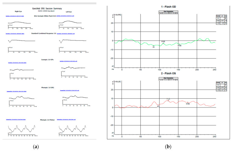
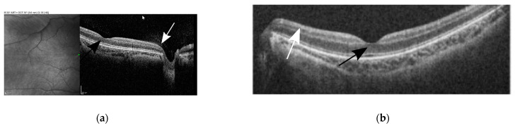
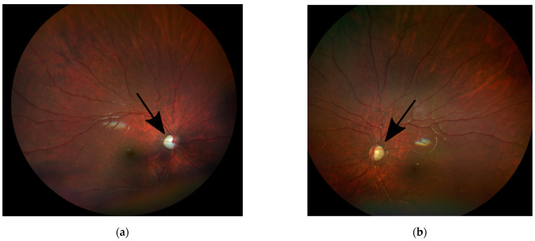
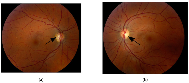
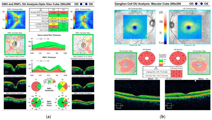
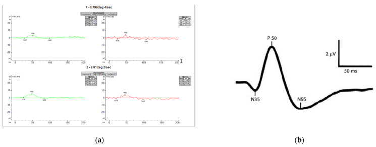
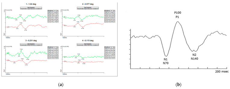
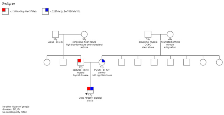
Dominant optic atrophy (DOA), MIM # 605290, is the most common hereditary optic neuropathy inherited in an autosomal dominant pattern. Clinically, it presents a progressive decrease in vision, central visual field defects, and retinal ganglion cell loss. A biallelic mode of inheritance causes syndromic DOA or Behr phenotype, MIM # 605290. This case report details a family with Biallelic Optic Atrophy 1 (OPA1). The proband is a child with a severe phenotype and two variants in the OPA1 gene. He presented with congenital nystagmus, progressive vision loss, and optic atrophy, as well as progressive ataxia, and was found to have two likely pathogenic variants in his OPA1 gene: c.2287del (p.Ser763Valfs*15) maternally inherited and c.1311A>G (p.lIle437Met) paternally inherited. The first variant is predicted to be pathogenic and likely to cause DOA. In contrast, the second is considered asymptomatic by itself but has been reported in patients with DOA phenotype and is presumed to act as a phenotypic modifier. On follow-up, he developed profound vision impairment, intractable seizures, and metabolic strokes. A literature review of reported biallelic OPA1-related Behr syndrome was performed. Twenty-one cases have been previously reported. All share an early-onset, severe ocular phenotype and systemic features, which seem to be the hallmark of the disease.

Keywords: Behr disease; biallelic OPA1; optic neuropathy.


Conflict of interest statement

The authors declare no conflict of interest.

Figures

Figure 2
Figure 2
Electrophysiology of the child (a) sedated full-field ERG using Burien Allen electrodes and the Vera system. Both dark and light-adapted waveforms and amplitudes were normal compared with our standards for sedated ERG (b) Awake flash VEP performed using a Dyagnosis system. Very low, almost non-recordable amplitudes bilaterally, right eye worse than left. Pick amplitudes for the P120 waves are 2.27 mV right eye and 5.27 mV left eye. Normal amplitudes are over 6 mV [24].
Figure 3
Figure 3
Optical coherence tomography (OCT) of the right (a) and left (b) macula of the child shows formed fovea in both eyes (black arrows) and a thin retinal neuron fiber layer (white arrows).
Figure 1
Figure 1
Fundus photos of the right (a) and left (b) eyes of the child showing diffuse optic disc pallor in both eyes (black arrows).
Figure 4
Figure 4
Fundus photos of the right (a) and left (b) eyes of the mother showing mild temporal optic disc pallor in both eyes (black arrows).
Figure 5
Figure 5
Optical coherence tomography (OCT) of the retinal nerve fiber layer (RNFL) (a) and ganglion cell layer (GCL) (b) of the mother. Red color represents thinning below normal values. Yellow color represents a moderate thinning. Green color represents normal values.
Figure 6
Figure 6
Pattern ERG (a) recordings of Patient’s mother showing normal P50 amplitudes indicating normal photoreceptor function and decreased N95 amplitudes (not below the baseline) indicating ganglion cell dysfunction. (b) normal pattern ERG waveforms-the P50-wave is the initial positive deflection originating from RGCs and outer retinal photoreceptor cells, namely the macular cones. The N95-wave is the negative deflection following the P50-wave that originates from the inner retina. This wave component reflects the RGC function. The asterisks * means the values are relative to the baseline (flat line) and that is why some are positive (p) and other negative (n).
Figure 7
Figure 7
Pattern VEP (a) recordings of Patient’s mother showing slightly decreased amplitudes more significant (b) normal pattern VEP waveforms showing the amplitude and latency to N70, P100 and N155 waves. The asterisks * means the values are relative to the baseline (flat line) and that is why some are positive (p) and other negative (n).
Figure 8
Figure 8
Complete family pedigree. Red colored is the paternally inherited variant and blue color is the maternally inherited variant.

References

    1. Lenaers G., Hamel C., Delettre C., Amati-Bonneau P., Procaccio V., Bonneau D., Reynier P., Milea D. Dominant optic atrophy. Orphanet J. Rare Dis. 2021;7:46. doi: 10.1186/1750-1172-7-46. - DOI - PMC - PubMed
    1. Patrick Y.W.M., Chinnery P.F. Dominant optic atrophy: Novel OPA1 mutations and revised prevalence estimates. Ophthalmology. 2013;120:1712–1712.e1. - PMC - PubMed
    1. Carelli V., Ross F.N.C., Sadun A.A. Mitochondrial dysfunction as a cause of optic neuropathies. Prog. Retin. Eye Res. 2004;23:53–89. doi: 10.1016/j.preteyeres.2003.10.003. - DOI - PubMed
    1. Olichon A., Guillou E., Delettre C., Landes T., Laetitia A.-P., Emorinea L.J., Mils V., Daloyau M., Hamel C., Patrizia A.B. Mitochondrial dynamics and disease, OPA1. Biochim. Et Biophys. Acta Mol. Cell Res. 2006;1763:500–509. doi: 10.1016/j.bbamcr.2006.04.003. - DOI - PubMed
    1. le Roux B., Lenaers G., Zanlonghi X., Patrizia A.B., Chabrun F., Foulonneau T., Caignard A., Leruez S., Gohier P., Procaccio V., et al. OPA1: 516 unique variants and 831 patients registered in an updated centralized Variome database. Orphanet J. Rare Dis. 2019;14:124. doi: 10.1186/s13023-019-1187-1. - DOI - PMC - PubMed

LinkOut - more resources